

Classification
Published online: 02 September 2026
Suggested citation
National Pressure Injury Advisory Panel, European Pressure Ulcer Advisory Panel and Pan Pacific Pressure Injury Alliance. Classification. In: Prevention and Treatment of Pressure Ulcers/Injuries: Clinical Practice Guideline. The International Guideline: Fourth Edition. Emily Haesler (Ed.). 2026. [cited: download date]. Available from: https://internationalguideline.com.
Introduction
A pressure injury (PI) is defined as localized damage to the skin and/or underlying tissue, as a result of pressure or pressure in combination with shear. PIs usually occur over bony prominences but may also be related to devices or other objects in close contact to the skin surface.Open wounds from various etiologies (e.g., venous ulcers, arterial ulcers, neuropathic ulcers, incontinence-associated dermatitis, skin tears and intertrigo) may appear similar to a pressure injury. The treatment of any wound begins with a comprehensive understanding of its etiology. Therefore, differentiating PIs from other wounds is the first step in assessing the wound and developing a treatment plan.
As with many other types of wounds, pressure injury terminology has been developed to describe the type of tissue damage. A PI classification system describes the extent of skin and tissue damage presenting as a pressure injury. Numerous classification systems have been developed and used over the years, informed by the evolving understanding of the etiology of PIs. Anatomical knowledge of the skin, subcutaneous fat, fascia and muscle layers (as well as supporting structures such as tendon, ligament and bone) is essential for accurate classification. The type and depth of tissue may vary depending on the anatomical site. Current classification systems are based on visual inspection of tissue and to some extent palpation for tissue consistency and temperature differences (see the guideline chapter on Skin and Tissue Assessment). Unfortunately, the true extent of tissue injury cannot always be detected with standard visual and tactile assessment. There are emerging assessment technologies that may improve diagnostic accuracy in the future. These are described in the guideline chapter on Advanced Technologies forSkin and Tissue Assessment.
This chapter will discuss differential diagnosis and classification of PIs. The chapter will provide an overview of classification systems that are commonly used around the world to identify the extent and type of tissue damage of presenting PIs. Terminology and classification systems continue to evolve as more research informs knowledge of pressure injury etiology. Throughout this guideline terminology from the International NPUAP/EPUAP Pressure Ulcer Classification System (2014) (1) is used to describe Category/Stage of PIs. However, the term ‘injury’ has been adopted in place of ‘ulcer’ due to increasing international recognition (2, 3). Tables 7and 8 in this chapter provide a crosswalk between terminologies used across different geographic and clinical settings.
Differential Diagnosis
CL1: Good Practice Statement
It is good practice to differentiate pressure injuries from other types of wounds based on etiology and clinical characteristics.
More information
Clinical question: How should pressure injuries be differentiated from other types of wounds?
Implementation considerations
Identify the most likely etiology of the wound. In the case of PIs, the injured area will usually be on a pressure loading surface in an individual with a history of mobility impairment or will be under a device (medical or other non-medical object).
Conduct a comprehensive assessment of the individual to inform differential diagnosis.
When applicable (e.g., particularly for foot and lower limb wounds), conduct a vascular assessment to inform differential diagnosis. The guideline chapter Preventing Heel Pressure Injuries includes guidance on performing vascular assessment.
Undertake and provide education on the etiology and clinical presentation of wounds commonly confused with PIs to enhance diagnostic accuracy.See Resources for specific guidance on differentiating PIs from other wounds and conditions, including:
o Category/Stage 2 PI versus incontinence associated dermatitis or intertriginous dermatitis in the pelvic area;
o PIs versus other types of chronic wounds in the lower extremities;
o Suspected deep tissue injury/deep tissue pressure injury (sDTI/DTPI) versus other conditions that cause purple discoloration.
Supporting information
Accurate determination of wound etiology is essential for the development of an appropriate and comprehensive treatment plan that addresses underlying PI risk factors. Accurate assessment of wounds and identification of their etiology is also important to inform the development and evaluation of PI quality improvement programs, to track PI incidence and prevalence, evaluate quality indicators and, in some geographic jurisdictions, to calculate reimbursement and/or monetary penalties applied to facilities (4).
Assessment of wound etiology is informed by the presentation of the wound, including its anatomical location and visual appearance. For example, wounds appearing over a bony prominence are more likely to be associated with pressure and shear, indicating a possible PI (4). Differentiation is also informed by a comprehensive assessment of the individual to identify comorbidities related to wound development and wound healing (e.g., diabetes mellitus, vascular disease or malnutrition) and to evaluate PI risk factors (e.g., exposure to shear forces, mobility level and continence). Environmental factors, particularly the presence of devices or equipment, are also a consideration in determining the etiology of the wound. A device-related PI (DRPI) usually conforms to the shape of the device, equipment or furniture that applied pressure to the skin and tissue (5).
Studies (6, 7, 8) have demonstrated that nurses with specialized knowledge in PIs or wounds, ostomy and continence care (WOC nurses) achieve high inter-rater agreement when classifying PIs. Highest levels of disagreement have been reported to relate to differentiation between moisture-associated dermatitis and PIs (7, 8). There is evidence that the accuracy of PI classification can be improved with education, and training (9, 10, 11, 12, 13, 14). This highlights the importance of ensuring access to appropriately trained health professionals and clinical leaders specializing in wound care. Those with specialized wound care training can review PI classifications determined by bedside nurses and validate or revise the classification as needed.
Resources
Resources to support differential diagnosis
Beeckman D, Barakat-Johnson M, Ahtiala M, Bernaerts K, Conway B, Dunk AM, Gannon R, Idensohn P, Santos V, Stenius M, Torra-Bou JE, Woodward S, Zobel K (2026) Incontinence associated dermatitis: moving prevention forward. Best Practice Update 2026. London: Wounds International. (Differential diagnosis and classification content starts on page 19).
Fletcher J, Beeckman D, Boyles A et al (2020) International Best Practice Recommendations: Prevention and management of moisture-associated skin damage (MASD).Wounds International.
Isoherranen K, Montero EC, Atkin L, Collier M, Høgh A, Ivory JD, Kirketerp-Møller K, Meaume S, Ryan H, Stuermer EK, Tiplica GS, Probst S. Lower Leg Ulcer Diagnosis & Principles of Treatment. Including Recommendations for Comprehensive Assessment and Referral Pathways. J Wound Management, 2023;24(2 Sup1):s1-76. (Contains an overview of multiple types and etiologies of lower leg ulcers including arterial ulcers and venous ulcers. Can be used to assist with a differential diagnosis of pressure injury of the lower extremity. The key differentiating feature for PI is pressure or pressure in combination with shear over a bony prominence or due to a medical device.)
Nair HK et al (2026) Implementation strategies for the prevention and management of diabetes-related foot disease. Wounds International. (Classification content starts on page 8. Also note multiple photos of diabetic foot ulcers).
Nokaneng E et al. Best practice recommendations for the prevention and management of skin tears in aged skin. Wounds International 2025. (Classification content on pages 5 and 19.)
Categorization of Pressure Injuries
CL2: Good Practice Statement
It is good practice to use a pressure injury classification system with clearly defined categories/stages to document the level of tissue loss. The classification system should include:
Category/Stage 1
Category/Stage 2
Category/Stage 3
Category/Stage 4
Unstageable
Suspected Deep Tissue Injury/Deep Tissue Pressure Injury.
Mucosal Membrane Pressure Injuries should not be categorized or staged.
More information
Clinical question: What are the recognized characteristics of each pressure injury category (i.e., Category/Stage 1 to 4, unstageable pressure injury and suspected deep tissue injury)?
Implementation considerations
Consistently use the same PI classification system to assess the PI.
Use a PI classification system to classify and document the level of tissue loss in a DRPI.
Do not use a PI classification system to describe tissue loss in a mucosal membrane PI.
Do not use a PI classification system to describe tissue loss in other types of wounds.
Reclassify sDTIs/DTPIs after full evolution based on the type of tissue seen at the base of the wound.
If debrided, unstageable PIs should be reclassified based on the type of tissue seen at the base of the wound and are usually a Category/Stage 3 or 4 PI. Do not debride stable heel PIs or PIs in other poorly perfused areas.
Additional implementation considerations for special populations
-
When classifying Category/Stage 1 PIs and sDTIs/DTPIs with intact skin in individuals with dark skin tones, rely on assessment of skin temperature, focal edema, change in tissue consistency, presence of skin pain and assessment findings from early detection technologies (if available) rather than identification of erythema. Assessment of these factors should also be included when evaluating extent of tissue involved in Category/Stage 2 to 4 PIs in individuals with dark skin tones (15, 16). See the guideline chapter Skin and Tissue Assessment.
Supporting information
PIs are classified according to the amount of visible tissue loss using a PI classification system. The use of a PI classification system:
Contributes to development of a PI prevention plan,
Informs the selection of PI treatments,
Improves communication between health professionals,
Allows for comparison of data between health services, and
Improves the methodological quality of PI research.
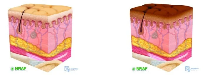
PI classification systems describe the extent of tissue involvement of a PI. Table 1 presents illustrations of healthy skin, and tissue in deeper structures, identifying five layers:
Epidermis
Dermis (papillary and reticular layers)
Adipose tissue
Muscle
Bone.
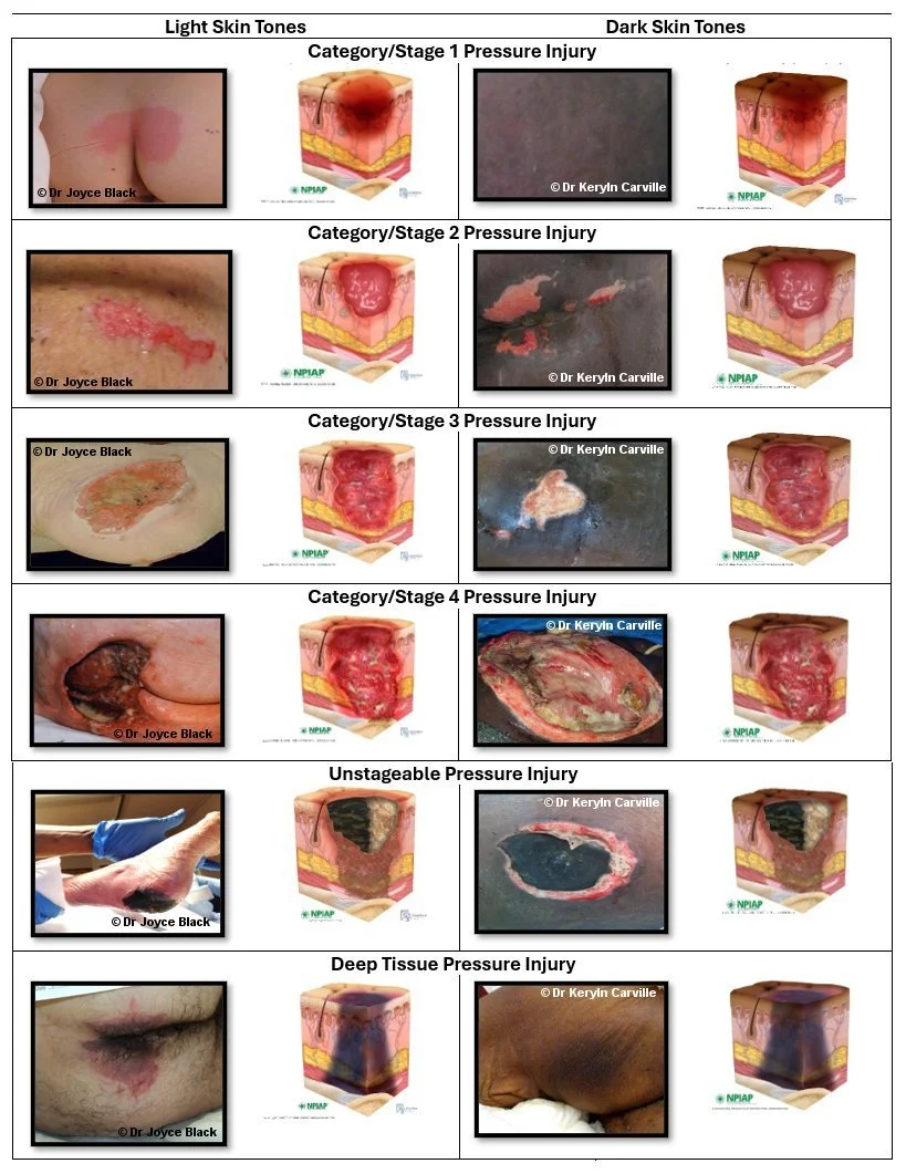
Table 3 presents photographs and illustrations of PI Categories/Stages, demonstrating the extent of tissue loss indicative of each Category/Stage.
Table 1: Illustrations of healthy skin (used with permission).
PI classification is based on the visual and palpatory identification of tissues including skin, subcutaneous fat, bone, muscle, tendon, and ligament. Necrotic tissue (eschar) and slough appear in full-thickness PIs. Granulation tissue becomes present as a full-thickness PI heals. In contrast, Category/Stage 2 PIs do not have necrotic tissue or slough and usually heal with epithelialization rather than granulation tissue. Healing tissues for full-thickness PIs include granulation tissue, epithelium and scar tissue.
PI depth varies by anatomical site, therefore relying on depth alone to determine whether a PI is Category/Stage 3 or 4 can be misleading. In anatomical locations with little adipose tissue (e.g., the bridge of the nose, the occiput, behind the ear, the sacrum, and the malleolus) shallow PIs can be Category/Stage 4. In contrast, in anatomical locations with greater adipose tissue (e.g., buttocks and ischium) a PI may be deep but not reach the muscle or bone and therefore, would be classified as a Category/Stage 3 or 4 PI.
The description of a PI should be supplemented with other findings. Indicating the exact anatomical location of the PI is important, including clearly identifying location over a bony prominence as applicable. Historical information, such as the conditions under which the PI developed, the history of prior treatment, and the trajectory of healing or non-healing of the PI (if known) should be documented and considered when evaluating the effectiveness of the treatment plan. See the guideline chapter on Supporting Pressure Injury Healing for specific parameters for assessing a PI (e.g., length, width, depth, exudate, types of tissue, and signs and symptoms of infection) and guidance for monitoring whether the PI is healing or stalled or deteriorating and therefore, requiring a change in the treatment plan.
Resources
Resources to assist with classification of pressure injuries
European Wound Management Association (EWMA) and Wound Australia: Pressure Injury Recurrence Toolkit
National Pressure Injury Advisory Panel (NPIAP): Evolution of Deep Tissue Pressure Injury
NPIAP: How to Inspect Darkly Pigmented Skin (DST) for Early Signs of Pressure Injury
NPIAP: Pressure Injury Train Modules 1-3: Staging, Differential Diagnosis and Prevention (Free to access after free registration).
CL3: Good Practice Statement
It is good practice to verify that there is accuracy and clinical agreement in pressure injury classification amongst the health professionals responsible for classifying an individual’s pressure injuries. Providing initial and ongoing education and clinical experience in pressure injury classification is essential to accurate pressure injury classification..
More information
Clinical questions: What is the interrater reliability, intrarater reliability and accuracy of pressure injury classification? What are strategies for improving and maintaining reliability and accuracy?
Numerous published studies have examined clinical agreement in PI categorization/staging and reported interrater reliability for various PI classification systems (see Table 5). These studies have either compared bedside evaluations of wounds or evaluated assessments of PIs based on photographs. Across studies, interrater reliability varied but was generally good to excellent for all classification scales. There appears to be limited variation in the reliability of assessment based on the observer/rater’s level of experience (8, 39), although in most of the reported studies, observers were registered/accredited nurses or health professionals with specific expertise in wound care and/or PIs.
Table 6 summarizes recent studies determining accuracy by comparing clinician classification of PIs to those of experts. Accuracy varies between studies and by category/stage. Unstageable, Category/Stage 3 and sDTI/DTPI seem to be particularly difficult to assess accurately (40, 41, 42, 43). Evidence suggests that AI assistance may improve accuracy, and this is likely to improve as AI-based systems for PI classification are developed more fully (42).
Supporting information
Table 5: Inter-rater reliability of categorization/staging using various pressure injury classification systems
| Classification system | Type of observation | Observers/raters | Interrater reliability unless otherwise stated |
|---|---|---|---|
| NPUAP classification system | Clinical assessment(44) | Nurses (n=180, n=591 observations) | κ=0.60 for Category/Stages 1 to 4 κ=0.61 for Category/Stages 2 to 4 |
| Clinical assessment(45) | WOCMs in hospitals (n=3) | κ=0.78, p<0.39 to 0.58, p<0.001 Agreement 55% to 62% |
|
| Photograph assessment(44) | Nurses (n=180, n=591 observations) | κ=0.69 for Category/Stages 1 to 4 | |
| Photograph assessment(46) | Academic nurses (n=3; 694 photos) | κ=0.84, p<0.001 to κ=0.94, p<0.001 | |
| EPUAP classification system 1999 | Photograph assessment(7) | RNs (n=20, n=56 photographs) | κ=0.80, p<0.01 |
| Photograph assessment(7) | PI nurses (n=17, n=56 photographs) | κ=0.78, p<0.01 | |
| Photograph assessment(8) | Nurses (n=473, n=56 photographs) | κ=0.41 to κ=0.50 Average agreement 55.6% |
|
| Photograph assessment(12) | Nurses and physicians (n=54, n=20) | κ=0.58 | |
| Photograph assessment(47) | Nurses working in a spinal cord injury setting (n=414, n=50 photos) | Agreement varied from 67& to 100% between Category/Stages | |
| Photograph assessment(7) | Wound researchers (n=7, n=56 photos) | κ=0.80, p<0.01 |
Table 6: Accuracy of categorization/staging in relation to expert categoris
| Classification system | Type of observation | Observers/raters | Accuracy in comparison to expert |
|---|---|---|---|
| NPUAP classification system | Photographic assessment(41) | Nurses (n=248, n=21 photographs) | 54.4% correct response rate for all. 54.4% correct response rate for all. |
| Photographic assessment(48) | Critical care nurses (n=2,778; 20 photographs)/td> | C45% correct response rate for all. 52.7% correct response rate for IAD 56.1% correct response rate - PI + IAD |
|
| Photograph assessment (including AI assistance)(42) | Dermatology residents (Derm. Res., n=8) Final year medical students (n=8) Hospital nurses (n=8) Database=3,098 images classified by experts |
Accuracy (image only):
|
|
| EPUAP classification system 1999 | Observation of registered nurses (RNs), reviewed by experts(40) | Observation of registered nurses (RNs), reviewed by experts(40) |
Percent of nurses' original categorization validated by experts:
|
| Photograph assessment(43) | Total (n=314) Nurses (n=199) Nursing students (n=115) 24 photographs with category validated by 5 experts. |
All participants.
|
Supporting Pressure Injury Categorization
Classifying Category/Stage 2 pressure injuries
Most Category/Stage 2 pressure injuries present as ‘a shallow open ulcer with a red, pink wound bed’. The red-pink color is consistent with tissue in the papillary dermis (top dermal layer, under epidermis) that is highly vascular and composed primarily of loose connective tissue. Category/Stage 2 can occasionally present as a shallow wound with yellow-white wound bed that can be confused with the slough of a Category/Stage 3 pressure injury. The yellow-white color is consistent with the tissue in the reticular layer of the dermis (bottom layer) that is composed of dense connective tissue(17) and is less vascular. The appearance of hair follicles and the relative depth of the wound can be used as differentiating features to avoid misclassification.
Table 2: Variations in Category/Stage 2 pressure injuries appearance (photographs used with permission)
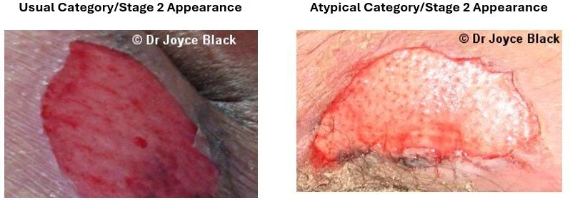
Table 3: Pressure injury Category/Stage: photographs and illustrations for light and dark skin tone (photographs used with permission).
Classifying Suspected Deep Tissue Pressure Injury/ Deep Tissue Pressure Injury
Due to its evolving nature, a sDTI /DTPI may be difficult to classify. It may appear as purple or maroon discoloration of intact skin or a blood-filled blister over a pressure loading surface. Evolution may include a thin blister over a dark wound bed or thin eschar. In some cases, the underlying tissue heals without any loss of the dermis or epidermis. Every sDTI/DTPI that progresses or is debrided (revealing deeper levels of tissue injury typical of Category/Stage 3 or 4 PI) should be reclassified as a Category/Stage 3 or 4 PI (1). Further discussion on differentiation of sDTI/DTPI from skin failure is included in the guideline chapter Research Priorities and Future Clinical Questions.
Visual inspection of the closed PIs (e.g., Category/Stage 1 or sDTI/DTPI with intact skin) may be augmented with an assessment of skin temperature using infrared thermography and focal edema using bioimpedance measurement devices, especially in individuals with medium and dark skin tones. As discussed in detail in the guideline chapter Skin and Tissue Assessment, areas of erythema are more difficult to identify and differentiate in medium and dark skin tones (18, 19, 20, 21, 22, 23, 24, 25), sometimes leading to a failure to detect Category/Stage 1 PI (18, 19, 26). Therefore, detecting localized heat using touch, skin thermometer or thermographic imaging; focal edema as detected by bioimpedance devices; changes in tissue consistency in relation to surrounding tissue and localized skin pain are all important indicators of pressure damage in medium and dark skin tones. Additionally, these tests might facilitate the diagnosis of a surrounding cellulitis in Category/Stage 2 or higher PIs in people with medium and dark skin tones.
The guideline chapters on Skin and Tissue Assessmentand Advanced Technologies for Skin and Tissue Assessment provide recommendations on assessment techniques (e.g., skin temperature, long wave infrared thermography with imaging, focal edema measurement and skin tone color charts) that can be used when assessing the skin and tissues and classifying PIs, in all skin tones. The guideline chapter on Supporting Pressure Injury Healingincludes clinical guidance on assessing PI-related pain.
Classifying healing, healed, and recurrent pressure injuries
As full-thickness PIs heal, the visible tissue(s) that initially supported the diagnosis of a Category/Stage 3 or 4 PI may be obscured by granulation tissue. Most experts advise against “downstaging” or “reverse staging”. Despite the numerical descriptors that identify levels of tissue damage for each stage, PIs do not progress from Category/Stage 1 through 4 and do not heal from Category/Stage 4 to 1. “Reverse staging from 4 to 3 to 2” would require the clinician to estimate the level of tissue replaced by granulation tissue. The science of wound healing does not support this approach. Granulation tissue does not replace damaged bone, muscle or subcutaneous fat. Most experts recommend that granulating PIs be reported as ‘healing Category/Stage 3 or 4’, depending on the original classification (if known).
A ‘healed pressure injury’ is one that is completely closed, fully epithelialized, covered completely with epithelial tissue, or resurfaced with new skin, even if the area continues to have some surface discoloration (27). This is a useful definition for clinical practice and as an endpoint for clinical studies; however, the healing process for full-thickness wounds continues for months to years after the wound has completely closed. The maturation/remodeling process continues with degradation and re-synthesis of extracellular matrix components to convert the type III collagen of granulation tissue to type I collagen and remodel the scar tissue. Even after full remodeling, the wound scar has only 80% the strength of the tissue prior to injury (28). This makes the area vulnerable to damage from pressure and shear.
Loss of skin integrity over areas of previously healed full thickness pressure injuries may be difficult to classify. The NPIAP recommends that these injuries be classified as ‘reopened, recurrent or new’ depending on the length of time since the previous pressure injury closed and the maturation of the scar tissue (29).
Classifying operative repairs of pressure injury
If a PI is surgically closed with a flap or graft, it should be coded as a surgical wound and not as a PI. If the flap or graft fails, continue to code it as a surgical wound until healed (27). Recommendations regarding classification of recurrent PI at healed flap/graft sites is unclear. Clinicians should consult and follow the policy of their local organization.
Classifying device-related pressure injuries
DRPIs result from medical devices, equipment, furniture and everyday objects that have applied pressure to the skin, either as an unintended consequence of their therapeutic use, or inadvertently due to unintended skin-device contact. When the PI is the result of a device designed and applied for diagnostic or therapeutic purposes it is referred to as a DRPI. The resultant PI generally conforms to the pattern or shape of the device (5). The term ‘device-related’ describes the etiology of the PI rather than its severity or extent of tissue loss (30). DRPIs should be staged using a recognized classification system, as for other PIs (30). See the guideline chapter Device-Related Pressure Injuries.
Classifying mucosal membrane pressure injuries
Mucosal membrane pressure injuries (MMPI) are pressure injuries of the moist membranes that line the respiratory, gastrointestinal and genitourinary tracts (31). MMPIs are primarily caused by medical devices (generally tubing and stabilization equipment) exerting sustained compressive and shear forces on the mucosa. In the mucosa of the respiratory tract (i.e., lips, mouth, nasal passages, etc.), PIs are typically caused by ventilation or feeding tubes and/or their stabilization equipment (31). Gastrointestinal tract PIs (32) and PIs of the genitourinary tract (i.e., penile, urethral, etc.)(33) are primarily caused by feeding tubes or ostomy appliances and catheters. In acute care, the most common causative devices have been shown to be endotracheal tubes (70.3%), urinary catheters (15.5%); gastric tubes (8.3%); nasal prongs (3.5%) and tracheostomy tubes (2.4%) (34).
Table 4: Mucosal membrane pressure injury (photographs used with permission).
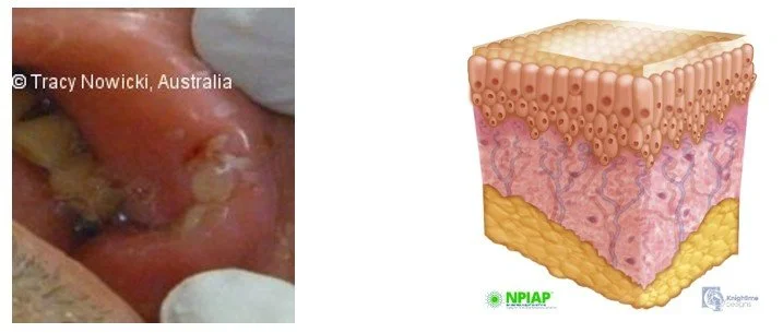
Classification systems for PIs of the skin and underlying tissues cannot be used to categorize MMPIs (30, 35). Where pressure is a significant factor in the etiology of the mucosal wound, it should still be considered a PI; however, it is inappropriate to use a PI classification system to categorize/stage. There are several factors that render classification systems designed for PIs of the skin inappropriate in the classification of MMPIs, including (35):
Non-blanchable erythema cannot be seen in mucous membranes. Shallow open mucosal ulcers indicating superficial tissue loss of the non-keratinized epithelium are so shallow that the naked eye cannot distinguish them from deeper, full thickness PIs.
Soft coagulum seen in MMPIs visually appears similar to slough that is often present in Category/Stage 3 PIs. However, this is actually a soft blood clot.
Exposed muscle is rarely visible in MMPIs.
There are efforts to develop a classification system for MMPIs (e.g., the Reaper Oral Mucosa Pressure Injury Scale [ROMPIS] (36, 37, 38)); however, no classification system for MMPI has demonstrated the level of reliability and validity necessary for recommending clinical use. The current recommendation is to continue to document MMPI but not assign a stage or category.
Education in pressure injury classification
Education underpins accurate and reliable PI classification. Numerous studies (9, 10, 11, 12, 13, 14, 49) provide evidence that health professional education increases accuracy of PI classification. Programs that have demonstrated success in improving classification skills have included various combinations of theoretical background information/didactic lectures (9, 12, 13, 14), practice by classifying PI photographs (9, 12, 13, 14), classification of PI videos (14), educational case studies (13), participation in monthly pressure injury risk, prevalence and incidence surveillance program (11), and clinical experience.(11, 43) A meta-analysis of six RCTs and two non-RCTs specifically explored the effectiveness of e-learning programs (49). The pooled standardized mean difference (SMD) for knowledge score and for skills in PI classification were 1.40 (95% CI 0.45 to 2.35) and 1.75 (95% CI 0.94 to 3.24) respectively. For the four studies reporting continuous outcomes, PI classification skills were significantly better for those completing e-learning when compared to usual education programs (pooled SMD 2.53, 95% CI 1.03 to 4.03, p < 0.001).
Artificial intelligence assisted pressure injury classification
There has been several key developments in the use of artificial intelligence (AI) in classifying PIs. Several AI systems can determine size and depth of a wound (50) and tissue types (51, 52). with a good degree of accuracy. Chao et al. (2025) (53) conducted a systematic review of 15 studies and a meta-analysis of eight studies exploring the accuracy of various methods of PI classification. Risk of bias varied between studies. Accuracy of clinician bedside classification was highly variable ranging between 16.65% and 71.63%. The meta-analysis involved 8 studies with 24 machine learning models classifying pressure injuries based on photographs verified by experts. Pooled sensitivity was 0.81 (95% CI 0.80 to 0.81, p < 0.0001); specificity was 0.87 (95% CI 0.86 to 0.87, p < 0.0001); AUROC was 0.93 (SE 0.02); and OR 20.48 (95% CI 15.37 to 27.27, p < 0.0001). When three methods of PI classification were examined (i.e., AI classification, human classification and human classification with AI assistance), AI classification (with AUROCs by category/stage ranging between 0.91 and 0.99) was slightly more accurate than human classification alone; however human classification improved significantly with AI assistance (42). The preferred model of the future may be human bedside evaluation with AI assistance. Nothing can replace bedside clinical assessment by a skilled clinician who can conduct a holistic and individualised assessment; however, AI analysis of photographs may detect some color or texture nuances undetectable by the human eye and serve as an important decision support tool. Ultimately, the validation and interpretation of findings is the responsibility of the clinician.
Pressure Injury Classification Systems
CL4: Good Practice Statement
It is good practice to consistently use the pressure injury classification system adopted by your healthcare system.
More information
Clinical questions: What are commonly recognized and used pressure injury classification systems and how do they relate to one another? Which classification system should be used?
Supporting Information
Supporting information
Generally, a healthcare system tends to adopt a single PI classification system. The system should align with the coding system used in their country to support reliable and accurate national data capture. Tables 7 and 8 present the most commonly used classification systems in Europe, the Pan Pacific and the US. These systems include those derived from classifications developed by the NPIAP in 1989, 2007 and 2016 (30, 54, 55); the NPUAP-EPUAP International Classification System which was adapted from the 2007 NPUAP System and published in the 2009, 2014, and 2019 International PI Guideline; and those developed by the World Health Organization (WHO) for international standards of disease reporting. In clinical settings, classification systems derived from NPIAP systems are most commonly used, while administrative coders usually adopt WHO-ICD classification systems. As noted in Tables 7 and 8, there are additional country-specific and setting-specific variations in terminology between classification systems. As our knowledge of PI etiology and the clinical assessment has advanced, continuous improvements have been made in classification systems. Current systems are similar, with minor differences in terminology and definitions. These systems are compared and contrasted below. Clinicians are encouraged to use the system adopted by their healthcare system to ensure consistency within their systems and comparability in reporting. However, one of the classification systems described in Tables 7 and 8 is recommended to ensure comparable data for meta-analysis and international reporting.
Resources
Resources for pressure injury staging
International Guideline classification tools (these classification systems may be reproduced for education purposes in full as they appear in the links i.e., the format, presentation, graphics and text remain unchanged):
Staging posters using the International NPUAP-EPUAP Staging System (2009, 2014, 2019):
Pressure Injury Classification System: Multicultural settings
Pressure Injury Classification System for Neonates and Children
Staging posters using the NPIAP Staging System (2016):
Crosswalks of classification systems
References
1. National Pressure Ulcer Advisory Panel (NPUAP), European Pressure Ulcer Advisory Panel (EPUAP), Pan Pacific Pressure Injury Alliance (PPPIA). Prevention and Treatment of Pressure Ulcers: Clinical Practice Guideline. Emily Haesler (Ed.) Cambridge Media: Osborne Park, WA; 2014
2. Ayello EA, Cordero GM, Sibbald RG. Survey Results from Canada and Some Latin America Countries: 2016 National Pressure Ulcer Advisory Panel Changes in Terminology and Definitions. Advances in Skin & Wound Care. 2017;30(2):71-6.
3. Ayello EA, Delmore B, Smart H, Sibbald RG. Survey results from the Philippines: NPUAP changes in pressure injury terminology and definitions. Advances in Skin & Wound Care. 2018;31(1):601-6.
4. Mahoney M, Rozenboom B, Doughty D, Smith H. Issues related to accurate classification of buttocks wounds. Journal of Wound, Ostomy and Continence Nursing. 2011;38(6):635-42.
5. Black J, Alves P, Brindle CT, Dealey C, Santamaria N, Call E, et al. Use of wound dressings to enhance prevention of pressure ulcers caused by medical devices. Int Wound J. 2015;12:322–7.
6. Hart S, Bergquist S, Gajewski B, Dunton N. Reliability testing of the National Database of Nursing Quality Indicators pressure ulcer indicator. Journal of Nursing Care Quality. 2006;21(3):256.
7. Defloor T, Schoonhoven L. Inter-rater reliability of the EPUAP pressure ulcer classification system using photographs. Journal of Clinical Nursing. 2004;13(8):952-9.
8. Defloor T, Schoonhoven L, Katrien V, Weststrate J, Myny D. Reliability of the European Pressure Ulcer Advisory Panel classification system. Journal of Advanced Nursing. 2006;54(2):189-98.
9. Briggs S-L. How accurate are RGNs in grading pressure ulcers? British Journal of Nursing. 2006;15(22):1230-4.
10. Young DL, Estocado N, Landers MR, Black J. A pilot study providing evidence for the validity of a new tool to improve assignment of national pressure ulcer advisory panel stage to pressure ulcers. Adv Skin Wound Care. 2011;24(4):168-75.
11. Tschannen D, McKay M, Steven M. Improving pressure ulcer staging accuracy through a nursing student experiential interventiom. J Nurs Educ. 2016;55(5):266-70.
12. Ham WHW, Schoonhoven L, Schuurmans MJ, Veugelers R, Leenen LPH. Pressure ulcer education improves interrater reliability, identification, and classification skills by emergency nurses and physicians. Journal of Emergency Nursing. 2015;41(1):43-51.
13. Lee YJ, Kim JY. Effects of pressure ulcer classification system education programme on knowledge and visual differential diagnostic ability of pressure ulcer classification and incontinence-associated dermatitis for clinical nurses in Korea. Int Wound J. 2016;13(1):26-32.
14. Beeckman D, Schoonhoven L, Fletcher J, Furtado K, Heyman H, Paquay L, et al. Pressure ulcers and incontinence-associated dermatitis: effectiveness of the Pressure Ulcer Classification education tool on classification by nurses. Qual Saf Health Care. 2010;19(5):e3.
15. Farid K, Winkelman C, Rizkala A, Jones K. Using temperature of pressure-related intact discolored areas of skin to detect deep tissue injury: An observational, retrospective, correlational study. Ostomy Wound Manage. 2012;58(8):20-31.
16. Bates-Jensen BM, McCreath HE, Pongquan V. Subepidermal moisture is associated with early pressure ulcer damage in nursing home residents with dark skin tones: Pilot findings. J Wound Ostomy Continence Nurs. 2009;36(3):277-84.
17. Brown TM, Krishnamurthy K. Histology, Dermis: National Library of Medicine and National Center for Biotechnology Information; 2022 [Available from: https://www.ncbi.nlm.nih.gov/books/NBK535346/#article-20394.s9].
18. Baumgarten M, Margolis D, van DC, Gruber-Baldini A, Hebel J, Zimmerman S, et al. Black/White differences in pressure ulcer incidence in nursing home residents. Journal of the American Geriatrics Society. 2004;52(8):1293-8.
19. VanGilder C, MacFarlane GD, Meyer S. Results of nine international pressure ulcer prevalence surveys: 1989 to 2005. Ostomy Wound Manage. 2008;54(2):40-54.
20. Meehan M. Multisite pressure ulcer prevalence survey. Decubitus. 1990;3(4):14-7.
21. Meehan M. National pressure ulcer prevalence survey. Adv Wound Care. 1994;7(3):27-30, 4, 6-8.
22. Lyder CH, Yu C, Stevenson D, Mangat R, Empleo-Frazier O, Emerling J, et al. Validating the Braden Scale for the prediction of pressure ulcer risk in blacks and Latino/Hispanic elders: a pilot study. Ostomy Wound Manage. 1998;44(3A Suppl):42S.
23. Lyder CH, Yu C, Emerling J, Mangat R, Stevenson D, Empleo Frazier O, et al. The Braden Scale for pressure ulcer risk: evaluating the predictive validity in Black and Latino/Hispanic elders. Appl Nurs Res. 1999;12(2):60-8.
24. Barczak CA, Barnett RI, Childs EJ, Bosley LM. Fourth national pressure ulcer prevalence survey. Adv Wound Care. 1997;10(4):18-26.
25. Baumgarten M, Margolis DJ, Selekof JL, Moye N, Jones PS, Shardell M. Validity of pressure ulcer diagnosis using digital photography. Wound Repair and Regeneration. 2009;17(2):287-90.
26. Rosen J, Mittal V, Degenholtz H, Castle N, Mulsant B, Nace D, et al. Pressure ulcer prevention in black and white nursing home residents: A QI initiative of enhanced ability, incentives, and management feedback. Adv Skin Wound Care. 2006;19(5):262-9.
27. Centers for Medicare & Medicaid Services. Long-Term Care Facility Resident Assessment Instrument 3.0 User's Manual. 2025. Version 1.20: Department of Health and Human Services USA,; 2025 [Available from: http://www.cms.gov/files/document/final-mds-3-0-rai-manual-v1-20-1-october-2025.pdf-1].
28. Wilkinson HN, Hardman MJ. Wound healing: cellular mechanisms and pathological outcomes. Open Biol. 2020;10(9):200223.
29. Tew C, Hettrick H, Holden-Mount S, Grigsby R, Rhodovi J, Moore L, et al. Recurring pressure ulcers: Identifying the definitions. A National Pressure Ulcer Advisory Panel white paper. Wound Repair and Regeneration. 2014;22(3):301-4.
30. National Pressure Ulcer Advisory Panel. NPUAP Pressure Injury Stages 2016 [Available from: https://cdn.ymaws.com/npuap.site-ym.com/resource/resmgr/npuap_pressure_injury_stages.pdf].
31. Coyer FM, Stotts NA, Blackman VS. A prospective window into medical device-related pressure ulcers in intensive care. Int Wound J. 2014;11(6):656-64.
32. Teno JM, Gozalo P, Mitchell SL, Kuo S, Fulton AT, Mor V. Feeding tubes and the prevention or healing of pressure ulcers. Arch Intern Med. 2012;172(9):697-701.
33. Bell MA. Severe indwelling urinary catheter-associated urethral erosion in four elderly men. Ostomy Wound Manage. 2010;56(12):36-9.
34. Fulbrook P, Lovegrove J, Butterworth J. Incidence and characteristics of hospital-acquired mucous membrane pressure injury: A five-year analysis. Journal of Clinical Nursing. 2023;32(13-14):3810-9.
35. National Pressure Ulcer Advisory Panel. Mucosal Pressure Ulcers: An NPUAP Position Statement NPUAP; 2008 [Available from: https://cdn.ymaws.com/npuap.site-ym.com/resource/resmgr/position_statements/mucosal_pressure_ulcer_posit.pdf].
36. Reaper S, Green C, Gupta S, Tiruvoipati R. Inter-rater reliability of the Reaper Oral Mucosa Pressure Injury Scale (ROMPIS): A novel scale for the assessment of the severity of pressure injuries to the mouth and oral mucosa. Aust Crit Care. 2017;30(3):167-71.
37. Fitzgerald S, McTier L, Whitehead C, Masters K, Wynne R. Inter-rater reliability of descriptors for the classification of mucosal pressure injury: A prospective cross-sectional study. Australian Critical Care. 2023;36(2):179-85.
38. Ozdemir E, Korkmaz F, Dereli N, Akyol M. Assessing Oral Mucosal Pressure Injuries in Intubated Intensive Care Patients: Adaptation and Reliability of the Reaper Oral Mucosa Pressure Injury Scale. Int J Nurs Pract. 2026;32(3):e70159.
39. Bååth C, Hall-Lord M-L, Idvall E, Wiberg-Hedman K, Wilde Larsson B. Interrater reliability using Modified Norton Scale, Pressure Ulcer Card, Short Form-Mini Nutritional Assessment by registered and enrolled nurses in clinical practice. Journal of Clinical Nursing. 2008;17(5):618-26.
40. Fulbrook P, Lovegrove J. Reporting accuracy of pressure injury categorisation in an acute tertiary hospital: A four-year analysis. Journal of clinical nursing. 2023;23.
41. Park S, Kim EJ, Lee SJ, Kim EJ, Lee JY, Hong JE. Knowledge and Visual Differentiation Ability of the Pressure Injury Classification System and Incontinence-Associated Dermatitis among Hospital Nurses: A Descriptive Study. Healthcare (Basel). 2024;12(2).
42. Kim J, Lee C, Choi S, Sung DI, Seo J, Na Lee Y, et al. Augmented Decision-Making in wound Care: Evaluating the clinical utility of a Deep-Learning model for pressure injury staging. Int J Med Inform. 2023;180:105266.
43. Rodriguez-Calero MA, Fullana-Matas A, Miró-Bonet R. Classification of Pressure Injury Stages and Skin Damage Photographs by RNs and Nursing Students in Spain: An Observational Comparison Study. J Wound Ostomy Continence Nurs. 2021;48(5):403-9.
44. Bergquist-Beringer S, Gajewski B, Dunton N, Klaus S. The reliability of the National Database of Nursing Quality Indicators pressure ulcer indicator: A triangulation approach. Journal of Nursing Care Quality. 2011;26(4):292-301.
45. Jesada EC, Warren JI, Goodman D, Iliuta RW, Thurkauf G, McLaughlin MK, et al. Staging and defining characteristics of pressure ulcers using photographs by staff nurses in acute care settings. Journal of Wound, Ostomy and Continence Nursing. 2013;40(2):150-6.
46. Karadaǧ A, Çakar V, Demir AS. An inter-assessor reliability study on the categorization and staging of pressure injuries. J Tissue Viability. 2024;33(4):786-91.
47. Sarhan F. Use of digital images in the assessment and treatment of pressure ulcers in patients with spinal injuries in community settings. Journal of Telemedicine and Telecare. 2010;16(4):207-10.
48. Liu H, Du A, Tian Y, Gao M, Ji S, Mi J, et al. The ability of critical care nurses to identify pressure injury and incontinence-associated dermatitis: A multicentre cross-sectional survey. Nursing Open. 2023;10(3):1556-64.
49. Ding Y, Qian J, Zhou Y, Zhang Y. Effect of e-learning program for improving nurse knowledge and practice towards managing pressure injuries: A systematic review and meta-analysis. Nurs Open. 2024;11(1):e2039.
50. Sahbudak G, Gunes U. Comparing Digital, Mobile and Three-Dimensional Methods in Pressure Injury Measurement: Agreement in Surface Area and Depth Assessments. J Clin Nurs. 2026;35(1):172-81.
51. Kimura Y, Ikuta K, Ohga M, Umeda R, Nakagaki M, Suyama Y, et al. Improving the Accuracy of Diagnostic Imaging using Artificial Intelligence: A Method for Assessing Necrotic Tissue in Pressure Injury. Yonago Acta Med. 2025;68(3):262-8.
52. Reifs Jiménez D, Casanova-Lozano L, Grau-Carrión S, Reig-Bolaño R. Artificial Intelligence Methods for Diagnostic and Decision-Making Assistance in Chronic Wounds: A Systematic Review. J Med Syst. 2025;49(1):29.
53. Chao Q, Pei J, Wei Y, Yang Z, Wang X, Du L, et al. Evaluation methods of pressure injury stages: A systematic review and meta-analysis. J Tissue Viability. 2025;34(3):100894.
54. No author listed. Pressure ulcers prevalence, cost and risk assessment: consensus development conference statement-The National Pressure Ulcer Advisory Panel. Decubitus. 1989;2(2):24-8.
55. Black J, Baharestani MM, Cuddigan J, Dorner B, Edsberg L, Langemo D, et al. National Pressure Ulcer Advisory Panel's updated pressure ulcer staging system. Adv Skin Wound Care. 2007;20(5):269-74.
56. World Health Organization. ICD-10 (Version 2016) 2016 [Available from: https://icd.who.int/browse10/2016/en.
57. World Health Organization (WHO). ICD-11 for Mortality and Morbidity Statistics (Version January 2026) https://icd.who.int/browse/2026-01/mms/en: WHO; 2026 [
58. National Centre for Classification in Health. International Statistical Classification of Diseases and Related Health Problems, Tenth Revision, Australian Modification. 11th ed2019.
59. World Health Organization. ICD-11 for Mortality and Morbidity Statistics (Version 04/2019) 2018 [Available from: https://icd.who.int/browse11/l-m/en.
60. Edsberg LE, Black JM, Goldberg M, McNichol L, Moore L, Sieggreen M. Revised National Pressure Ulcer Advisory Panel Pressure Injury Staging System: Revised Pressure Injury Staging System. Journal of Wound, Ostomy and Continence Nursing. 2016;43(6):585-97.
61. Centers for Medicare & Medicaid Services. Long-Term Care Facility Resident Assessment Instrument 3.0 User's Manual. 2019. Version 1.17:129. https://downloads.cms.gov/files/mds-3.0-rai-manual-v1.17.1_october_2019.pdf; 2019.
62. Centers for Disease Control and Prevention. ICD-10-CM Official Guidelines for Coding and Reporting - FY 2026 https://ftp.cdc.gov/pub/Health_Statistics/NCHS/Publications/ICD10CM/2026-update/ICD-10-CM%20April%201%202026%20Guidelines%20Final.pdf2026