

Pressure Injury Risk
Published online: 06 September 2026
Suggested citation
National Pressure Injury Advisory Panel, European Pressure Ulcer Advisory Panel and Pan Pacific Pressure Injury Alliance. Pressure Injury Risk. In: Prevention and Treatment of Pressure Ulcers/Injuries: Clinical Practice Guideline. The International Guideline: Fourth Edition. Emily Haesler (Ed.). 2026. [cited: download date]. Available from: https://internationalguideline.com.
Introduction
Risk assessment aims to identify individuals with characteristics that increase the probability/likelihood of PI development. Risk assessment is a central component of clinical practice and a necessary initial step aimed at identifying individuals susceptible to pressure injuries (PIs). Assessment of PI risk should then inform the development and implementation of an individualized management plan to mitigate modifiable risk factors and prevent PI development. Both modifiable and non-modifiable risk factors should be included in a full assessment of PI risk; however, PI prevention focuses on modifiable risk. Non-modifiable risk factors should be noted as they increase the probability of PI development but are not amenable to prevention (e.g., age or existing PI).
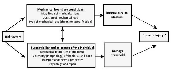
As detailed in Figure 1, individuals who are at high risk of PIs are those characterized by multiple risk factors that affect both:
Exposure to damaging mechanical boundary conditions (i.e., the type, magnitude, time and duration of the mechanical load), and
The susceptibility and tolerance of the individual (i.e., mechanical properties, geometry, physiology and repair, and transport and thermal properties of the skin and tissues).
PIs occur when the magnitude and duration of mechanical loads exceed the individual’s tissue tolerance (i.e., the damage threshold is exceeded).
Figure 1: Factors influencing the susceptibility of an individual for developing pressure injuries (Adapted from Oomens (1) by Coleman et. al. (2) reproduced with permission)
General Risk Factors and Their Assessment
Clinical question: What factors put individuals at risk for pressure injury development?
Decades of epidemiological studies have identified a potentially overwhelming number of risk factors for PIs. In evaluating the It is helpful the large volume of data on individual risk factors, it is helpful to consider common domains or categories of risk and whether specific risk factors affect mechanical boundary conditions or susceptibility or tolerance of the individual or both.
Table 1 provides an overview of the independent risk factors considered in published multi-variable modelling according to the key domain into which they can be categorized (2, 3). Table 2 includes examples of individual risk factors that are identifiable with clinical examination for each risk factor domain. This is not intended as an exhaustive list of risk factors or supporting evidence. Individual risk factors characteristic of each risk domain may vary between populations and settings. In addition to statistical significance in a multi-variable model (often using logistic regression), a risk factor should precede the disease, have a plausible physiological link to the etiology of PIs and be clinically significant.
Population specific risk factors are discussed in the guideline Chapter Pressure Injury Risk in Specific Populations.
Table 1: Major risk factor domains and their influence on components of the conceptual framework
| Risk factor domains | Mechanical boundary conditions(MBC) | Susceptibility and tolerance of the individual (ST) | Examples of individual risk factors identifiable in clinical setting |
|---|---|---|---|
| Activity and mobility limitations |
|
||
| Skin and tissue status |
|
||
| Perfusion, circulation and oxygenation factors |
|
||
| Nutrition indicators |
|
||
| Moisture |
|
||
| Body temperature |
| ||
| Sensory perception limitations: local or systemic |
|
||
| Blood markers |
|
||
| General health status |
|
||
| Demographic Variables: Extremes in age |
|
||
| Other Demographic variables |
|
||
| Medical devices |
|
RISK1: Good Practice Statement
It is good practice to conduct a pressure injury risk screening as soon as possible after admission to the care service and periodically thereafter to identify individuals at risk of developing pressure injuries. Rescreening should occur with any significant worsening in the individual’s condition (Step 1).
Clarifiers:
Screening should quickly and accurately identify individuals who are likely to be at risk.
At a minimum, screening should include a reliable measure of mobility or activity limitations, presence of medical devices and existing pressure injuries.
More information
Clinical question: What are the steps of pressure injury risk assessment and risk-based prevention planning?
RISK2: Good Practice Statement
It is good practice to conduct a full pressure injury risk assessment as guided by the screening outcome after admission, periodically and after any worsening in condition. Any individuals screened as “likely at risk” should have a full pressure injury risk assessment (Step 2).
Clarifiers:
A full pressure injury risk assessment should:
Use a structured approach.
Include a comprehensive skin assessment.
Supplement use of standardized risk assessment tools/risk scales (if used) with assessment of additional risk factors relevant to the population and setting.
Interpret the assessment outcomes using clinical judgment.
Document the risk assessment findings.
Examples of structured, full pressure injury risk assessments may include:
1. A summary of relevant evidence-based risk factors.
2. Quantitative risk assessment tools.
3. Qualitative risk assessment tool(s).
4. Artificial intelligence and machine learning based risk assessment (under development).
RISK3: Good Practice Statement
It is good practice to develop and implement a risk-based prevention plan for individuals identified as being at risk of developing pressure injuries (Step 3).
Clarifier:
Examples of prevention planning strategies include but are not limited to:
1. Generic care bundles: A collection/protocol of three or more generic interventions that apply to most at-risk individuals.
2. Risk-based: Interventions or bundles of interventions are explicitly driven by risk assessment tool items, subscales or specific individual risk factors.
3. Clinical algorithms: Clinical algorithms guide clinicians through a series of evidence-based interventions appropriate to the individual’s risk profile and health status.
RISK4: Good Practice Statement
It is good practice to use clinical judgment at each step of the risk assessment and prevention planning process.
RISK5: Good Practice Statement
It is good practice to document ongoing risk assessments and prevention plans.
Implementation considerations
Risk screening
Use a structured approach AND clinical judgment.
Risk screening should occur as soon as possible (i.e., at first contact with the health professional) (Step 1).
Screen for risk factors within your population that will quickly and accurately identify those likely at risk who should undergo full risk assessment.
At a minimum, include a reliable measure of mobility or activity limitations, presence of medical devices and existing pressure injuries.
Screening is most useful in low risk or mixed risk populations (73).
In high-risk populations where most individuals are obviously at risk (e.g., critical care or SCI), risk screening is unnecessary (73). In these situations, proceed directly to full risk assessment (Step 2).
Periodically monitor PI outcomes in the clinical population in relation to screening. If risk screening is not highly sensitive in the clinical context, and especially if it misses individuals who later develop PIs (false negative screen) then re-evaluate the risk factors selected for screening and the processes used for screening.
Full risk assessment
Use a structured approach together with clinical judgment. Examples include:
o A summary of relevant evidence-based risk factors.
o Quantitative risk assessment tools.
o Qualitative risk assessment tools.
Include a comprehensive skin assessment as a part of all risk assessments (see the guideline chapter Skin and Tissue Assessment).
Ensure all domains of risk are represented in the assessment. Table 1 outlines specific risk factors within each domain.
Supplement risk assessment tools and other structured approaches with additional risk factors as needed, particularly those specific to the population.
Ensure that selected risk factors are relevant and can be accurately assessed.
Risk assessment tools should be supported by reliability and validity testing (see Table 3). Self-developed tools usually do not have sufficient reliability and validity testing to support use in clinical practice.
Do not use total scores from a risk assessment tool as the sole basis for prevention planning. If a quantitative risk scale is applied, use tool subscales as a basis for risk-specific prevention planning.
Cut-off scores have been used in some countries to establish at-risk status versus not-at-risk status. Even if an individual is considered not-at-risk based on the cut-off score, activity-mobility deficits override cut-off-based decisions. If using a quantitative risk assessment tool, revise the definition of at-risk to accept a cut-off score in the at-risk range or to include any deficits in mobility-activity.
Risk-based prevention planning and implementation
Use a structured approach together with clinical judgment. Examples are discussed in this guideline chapter.
Select the approach that is most effective for the population.
Be aware of the strengths and weaknesses of each approach. For example, a generic care bundle may be appropriate for most individuals but a process is required for identifying very high-risk individuals and circumstances that may require an expanded or tailored approach.
Regardless of the approach selected for prevention planning and implementation:
o Measures should be planned and implemented taking individual preferences, self-care capacity and treatment goals into account.
o Discuss the PI prevention plan with the individual, their carers and all involved health professionals.
o Document compliance with plan.
o Revise plan with changes in medical condition and risk status.
Supporting information
For practical reasons, it is recommended that risk assessment and prevention planning is organized into three steps:
Step 1: Risk screening to quickly identify individuals who are likely at risk of getting a pressure injury (73),
followed by:
Step 2: A full pressure injury risk assessment in those individuals screened as being likely at risk (73),
followed by:
Step 3: Risk-based prevention planning and implementation.
These steps are illustrated in Figure 2.
The 3-Step Process for Risk Assessment and Prevention
Risk assessment and prevention planning is organized into three steps, as outlined in Figure 2, and discussed below.
Figure 2: Three-step process for risk assessment and prevention

Step 1: Risk screening
The first step, PI risk screening, aims to rapidly identify with a minimum of diagnostic effort those individuals admitted to any health care service (e.g., tertiary hospital, aged care facility, home care agency, rehabilitation facility, etc.) who are likely to be at risk of developing a PI (73). Thus, the main purpose of screening is to identify those individuals for whom PI risk cannot be ruled out instantly, thus prompting the need for a full risk assessment. Undertaking risk screening should help target resources to those individuals in need of a full risk assessment and preventive interventions, but should also safeguard that all individuals who are "likely at PI risk” are identified early.
The PI risk screening should follow a structured and replicable approach that considers relevant PI risk factors in the target population, the local health care infrastructure and procedures, and the training and scope of practice of the health care team.
To satisfy the screening purposes, screening must rely on a small number of highly predictive PI risk (i.e., risk factors that identify individuals with a high degree of likelihood of PI development). The major risk factors recommended for risk assessment in this guideline chapter provide a theoretically and empirically justified reference framework for PI risk screening. However, not all identified risk factors are equally predictive in all target populations. Risk factors that are highly prevalent in a specific population of interest do not help distinguish between individuals likely to be at PI risk and those likely not at PI risk.
The screening should only consider factors supported by a high level of evidence that have the potential to accurately identify individuals likely to be at PI risk in the population of interest. The primary cause of PI is pressure or pressure in combination with shear. Thus, pressure is a necessary condition for PI development. Therefore, risk factors associated with increased pressure and shear are excellent choices for risk screening. These include deficit in mobility/activity, shear with movement and presence of a medical device. Regardless of age or clinical setting, an individual should be considered at risk of a PI as soon as a device has been applied. Presence of an existing PI or history of a PI should also indicate an individual likely to be at PI risk.
There are also populations who may not require any formal screening because the presence of significant PI risk factors is associated with their reason for admission. For example, in critically ill individuals, premature/critically ill neonates, or individuals with SCI, several major risk factors are so obviously present and highly prevalent that any formal screening is superfluous or can be regarded as being automatically completed. In this case, the individual can be considered as having ‘at risk’ status at admission and a full PI risk assessment is required (73).
As the PI risk screening must be conducted rapidly, the process should rely on easily accessible information related to the individual’s health history and current health status. Information can be attained the individual themselves, or from their formal health records, informal carers or other health professionals. For example, existing mobility and activity limitations may be observable directly at the first contact or inferred from existing information on the individual’s needs for assistance. Information on the individual’s skin status and other major risk domains may be taken from health records or admission papers.
The outcome of PI risk screening is usually dichotomous (i.e., it indicates at least one highly predictive risk factor is present or that the highly predictive risk factor/s is not present). Individuals should be assumed as being at pressure injury risk if the screening results indicate that any of the risk factors included in the risk screening are present (74). Existing mobility or activity limitations, medical devices or existing pressure injuries should always be regarded as an indication of an “at risk” status (74, 75).
A PI risk screening should be conducted as soon as possible (i.e., at first contact with the health professional) or at first visit in community settings. Health services may wish to specify a specific time frame in which this first assessment must be conducted, and/or a frequency of reassessment. In individuals screened as likely not at PI risk, the screening should be repeated as soon as the risk exposure has increased or is likely to increase due to changes in the individual’s health condition or treatment (e.g., surgery). In individuals screened as being “likely at risk”, a full PI risk assessment should be undertaken immediately.
Step 2: Full risk assessment
A full PI risk assessment aims to thoroughly examine an individual’s risk exposure and identify modifiable risk factors. For this in-depth evaluation, all major risk factor domains recommended in this guideline chapter should be considered (see Table 1). Individual risk factors from each domain can be sufficiently broad to cover most populations or can be more specific to the population. For example:
Broad to cover most populations: activity status operationally defined within an ordinal scale such as bedrest → chairbound → walks with assistance → or walks independently, versus
Population specific: spinal cord injury as a clinical indicator of activity status.
Risk assessment identifies both modifiable and non-modifiable PI risk factors to provide an overall indication of the individual’s risk status, or “probability of developing a PI”. If the full assessment does not confirm the ‘likely at risk’ assumption indicated by the screening, individuals should be screened again as necessary. Rescreening should occur if the risk exposure has likely increased or is likely to increase due to changes in the individual’s health status or treatment (e.g., surgery). The presence and impact of each factor on the individual’s risk exposure should be evaluated by means of deliberate, comprehensive risk assessment methods. Full PI risk assessments should be repeated for individuals at-risk with every change in their condition or treatment. This ensures that any new modifiable risk factors are addressed in Step 3.
As noted in the guideline chapter on Skin and Tissue Assessment, a comprehensive skin assessment should be part of every full PI risk assessment. Skin and risk assessment are inextricably linked. As noted earlier in this chapter, there is epidemiological evidence that alterations in skin status (specifically the presence of an existing PI) are associated with development of new PIs, making skin assessment an essential component of any risk assessment. Additionally, PI risk factors such as skin moisture and pain at pressure points can be identified in a skin and tissue assessment and addressed in an individualized PI prevention plan.
Step 3: Risk-based prevention planning and implementation
When an individual is confirmed as being at risk of PI development, a prevention program that aims to minimize the impact of the identified modifiable risk factors should be developed. Risk assessment should naturally flow into prevention. Although a full risk assessment identifies both modifiable and non-modifiable risk factors, preventive interventions can only address modifiable risks. Non-modifiable risk factors should be considered in the overall assessment of the individual’s likelihood of PI development and may prompt more aggressive mitigation of modifiable risk factors in individuals with a high burden of non-modifiable risk factors. The rationale and measures of care should be explained to and agreed with the individual, and the agreed plan of care should be documented. Further discussion of risk-based PI prevention planning is below.
Documentation
Accurate ongoing documentation of risk assessments and prevention plans is essential. Documentation of risk assessments ensures communication within the multidisciplinary team, provides evidence that care planning is appropriate, and serves as a benchmark for monitoring the individual’s progress (77, 78).
Structured Approaches to Full Risk Assessment
Examples of structured approaches to full risk assessment include:
Summary of relevant evidence-based risk factors.
Quantitative risk assessment tools.
Qualitative risk assessment tools.
Artificial intelligence (AI) and machine learning (ML) based risk assessment (under development).
Summary of relevant evidence-based risk factors
Some local and national protocols identify relevant evidence-based risk factors that guide health professionals as they use their best-informed, well-reflected, clinical judgment to conduct a full risk assessment (73). The health professionals completes their risk assessment by utilizing patient history, conditions-specific tools, clinical/diagnostic information and their clinical judgment. Additional information on this approach can be found in national policies on PI risk assessment in Australia and Germany (73, 79, 80). The focus is on providing the best evidence possible to support clinical judgment.
Selecting relevant evidence-based risk factors can be challenging. Hundreds of risk factors have been identified through multi-variable analyses in epidemiological studies of PI risk. Selection of risk factors requires expertise in evaluating the research, as well as clinical expertise in determining which risk factors are meaningful in various clinical settings, as well as which factors can be reliably assessed. Reliability and validity data for this method are not readily available. This approach has been generally evaluated as PI prevalence and incidence rates. Rates are usually stable or improved (73). Investigations of the clinical flow from “this risk assessment strategy to risk-based prevention planning to reductions in pressure injury rates” have not been conducted. The advantage of this approach is that it allows flexibility in applying evidence-based risk factor knowledge to clinical decision-making. Its success in reducing PIs relies on the application of the clinical judgment of bedside health professionals to the full risk assessment process.
Clinical questions: What types of risk assessment tool are available? Which risk factor domains are represented in each tool? Which domains are missing from the tool and require consideration of additional risk factors for a full risk assessment?
Table 2includes a list of common risk assessment tools, including information on which risk domains they do and do not cover.
Quantitative risk assessment tools
Standardized risk assessment tools/scales with numerical scoring have been developed to operationally define the various domains of PI risk. Rather than summarize the specific risk factors from epidemiological studies, a quantitative risk assessment tool seeks to broadly identify the risk domains using clinical indicators or factors that can be easily assessed. For example, to assess the PI risk associated with moisture, a tool may include a descriptor of how often the individual is moist and requires skin care and a change in bed linen, in contrast to a list of factors such as urinary, fecal or dual incontinence. Risk assessment tools are often described as heuristic tools (81). When faced with an overwhelming amount of data (e.g., hundreds of risk factors), heuristics can create mental shortcuts to facilitate quicker decision-making. Substantial thought goes into creating a useful heuristic strategy. In the case of PI risk assessment, the hundreds of risk factors identified from multi-variable analyses should be analysed and synthesized to create a heuristic strategy that is pragmatic, if not perfect (82).
Subscale and total scores
Most risk assessment tools contain subscales that capture one or more of the risk domains (see Table 1). Subscales often use an ordinal scoring system to approximate the degree of risk within the domain. Subscale scores can be added for a total score on a risk tool. However, most experts caution against using a total score as the sole basis for prevention. Closer examination of the subscale scores to gain an appreciation for the type and degree of risk within each domain or subscale is recommended. There is often an emphasis on identification of modifiable risk factors that can be used as a basis for a PI prevention plan. A risk assessment tool rarely covers all domains of risk (see Table 2). Most experts recommend that clinicians examine additional risk factors and use clinical judgment when assessing risk and planning prevention.
General risk assessment tools
Risk assessment tools may be general in nature; for example, those used across general adult populations (e.g., Braden Scale (83-85), Norton Scale (86), and Waterlow Scale (87, 88)). Many PI risk assessment tools have been modified in an attempt to improve their performance in specific clinical settings. Multiple studies have investigated the predictive validity of different risk assessment tools, with varied results based on the population being studied (89-92).
Population-specific risk assessment tools
There is an ever-increasing trend toward developing risk assessment tools tailored to a specific population, including but not limited to infants and children (91-94), individuals in the operating room (7, 95), individuals in critical care (96-105), and individuals with SCIs (106-108). A recent umbrella review of systematic review identified 124 risk prediction tools (109). These tools are discussed in more detail in the guideline chapter Pressure Injury Risk in Specific Populations.
Reliability and validity considerations
A carefully designed and validated risk assessment tool should:
include as many relevant, evidence-based risk domains as possible,
be consistent with our understanding of etiology,
provide clear, unambiguous descriptions of clinical assessments that accurately capture risk, and
achieve results that can be reliably reproduceable by clinicians with minimal training.
As new risk assessment tools are developed, they should undergo:
content validation in relation to the conceptual framework of PI etiology,
reliability and validity testing, and
predictive validity testing (e.g., sensitivity, specificity, area under the receiver operator curve) to evaluate whether components of the scale accurately “predict” PI development.
Kottner and Coleman (2023) (110) identify specific phases of clinical prognostic PI risk research. The most frequently used risk assessment tools at least partially address the initial criteria for tool development (e.g., epidemiological evidence supporting risk factors, model development and testing) but generally fail to answer the question, “Does the use of the prognostic factor/model lead to better health outcomes?”
The effect of assessment tools on pressure injury incidence
A Cochrane systematic review (111) identified two low quality randomized controlled trials (RCTs) evaluating the effect of risk assessment tools on PI incidence. There was no significant difference between risk assessment and clinical judgment alone. It was unclear whether the results of the risk assessments were used as a basis for developing and implementing a risk-based prevention plan. For any risk assessment tool to influence PI outcomes, the clinician must act on the assessment results. Individuals with a higher level of risk tend to have more preventive interventions (112-113) Before-after and quality improvement studies have demonstrated a decrease in PI incidence when clinicians use risk assessment tool results (i.e., risk levels determined by total or subscale scores) as a basis for PI preventive care (114-118).
Lovegrove et. al. (2021) (119) conducted a systematic review examining this question by focusing on whether the risk assessment conclusion (i.e., level of risk) and prescribed preventive interventions differed between a health professional using a structured PI assessment tool compared to relying on their clinical judgment. The authors noted that clinical judgment is poorly defined and conceptualized in the literature making comparisons of risk assessment outcomes between the two approaches difficult. Evidence supporting the effectiveness of clinical judgment alone is not available. The link between risk assessment and preventive interventions is not optimal, regardless of risk assessment method (78, 112). Until that intermediate link is strengthened in clinical practice, an accurate evaluation of the effects of any risk assessment method on PI incidence cannot be performed (120).
Qualitative risk assessment tools
The Pressure Ulcer Risk Primary or Secondary Evaluation Tool (PURPOSE-T) is a qualitative risk assessment guide based on a systematic review of risk factor literature completed in 2013 (2) and the input of clinical nurse and patient caregivers (121). It contains a screening phase with progression to full risk assessment based on screening results. Multiple risk factors in various domains must be considered by the health professional, highlighting the need for clinicians to have comprehensive education and the skills to apply clinical judgment and decision-making. Through a series of color-coded checked/ticked boxes, the provider is led to one of three conclusions:
no PI and not currently at risk,
no PI but at risk and requires primary prevention, or
Presence of a Category/Stage 1 PI or greater, or scarring from a previous PI, that requires secondary prevention/treatment.
For PI prevention, PURPOSE-T refers health professionals to a care bundle with generic prevention strategies such as SSKIN (Surface, Skin inspection, Kinetics/keep moving, Incontinence/moisture, Nutrition/hydration) (121-122).
PURPOSE-T has been evaluated with positive results for content validity, feasibility (123), acceptability (124), usability, interrater and test-retest reliability, and both convergent and known groups validity (121, 125). Coleman (2018) (121) documented interrater reliability (i.e., simple kappa = 0.71; weighted kappa = 0.76) and test-retest reliability (i.e., 92% for three outcomes; 95.8% for a dichotomized outcome of at “risk or not at risk”) (121). There are no numerical scores in a qualitative risk assessment tool; therefore, more detailed, parametric statistical analyses have not been performed. A 2 x 2 table could be constructed to document whether subjects were considered at-risk (yes-no) versus PI present on follow-up (yes-no). This would allow calculation of some predictive validity measures (e.g., sensitivity, specificity, area under receiver operator curve). To our knowledge, this analysis has not been conducted and published.
Additionally, there are no RCTs linking PURPOSE-T use to lower PI incidence. Seton and colleagues (2025) (129) conducted a cross-sectional study comparing PI point prevalence before and after PURPOSE-T implementation. Prevalence rates were similar between before and after introduction of PURPOSE-T; however, more preventive interventions were implemented as a result of the tool use, and there was a decrease in the prevalence of Category/Stage 3 and 4 PIs following PURPOSE-T implementation.
Assessment Tool Selection and Interpretation of Results
Clinical questions: Which risk assessment tools have the best predictive validity? Which risk assessment tools are most appropriate to my patient population and also identify modifiable risk factors for preventive action?
While it is important to determine how well a tool predicts PIs in a controlled research design, the term prediction is less useful in clinical practice. If a health professional uses a tool subscale as a basis for PI prevention, the interventions should prevent the predicted PI; thereby lowering the risk tool’s predictive validity. In the clinical context, it is more useful to consider risk assessment tools to be a measure of the “likelihood” of PI development for a specific individual at a point in time, rather than predictive of an outcome. Both Braden (2012) and Norton encourage clinicians to consider risk assessment tools as primarily an assessment tool rather than a prediction tool (130). Table 2 includes a list of common risk assessment tools, including information on which risk domains they do and do not cover.
Validity refers to the degree to which a tool measures what it claims to measure. Of the many types of validity (e.g., content, construct and criterion), ‘predictive validity’ has received the most attention in relation to risk assessment tools. Rather than focus on the degree to which these tools accurately measure risk factors such as mobility, activity and skin moisture, we have focused on the degree to which they predict a future event (i.e., pressure injury development).
A major problem identified in the literature in establishing predictive validity of risk assessment tools is that preventive interventions are initiated in most studies, and these will impact upon the performance of the tool. Studies of predictive validity are prognostic (estimating the likelihood of a future problem) rather than diagnostic (identifying an existing problem). Despite these constraints, most studies of predictive validity report some statistical estimates of likelihood associated with each prognostic method. These include (definitions included in glossary):
Sensitivity.
Specificity.
Positive likelihood ratio (PLR).
Negative likelihood ratio (NLR).
AUROC curves.
Relative risk (RR).
Diagnostic Odds Ratio (DOR).
Although these measures are imperfect, they provide some insight into the predictive validity of risk assessment tools, especially in the context of the intervening preventive interventions. If selecting a risk tool for use in a specific population, predictive validity testing provides some guidance. However, this guidance should be balanced with the appropriateness of the tool for your patient population and its ability to identify modifiable risk factors for mitigation. As illustrated in Table 2, no risk assessment tool contains a full list of potential risk factors and should be supplemented with additional risk factors as needed.
The AUROC provides the best balance of sensitivity and specificity and an indication of the overall performance of a test. An AUROC ≥ 0.9 is considered excellent, 0.8 to 0.9 is considered good and 0.7 to 0.8 is considered fair. The AUROC for most PI risk assessment tools is in the fair range because concurrent preventive interventions interfere with a true and accurate picture of prediction.
Table 3 provides an overview of published psychometric qualities of major PI risk assessment tools to assist health professionals to selection a tool most likely to achieve good predictive validity in the relevant patient population. The data recorded in Table 3 was extracted from the most recent systematic reviews with meta-analysis. When a recent meta-analysis was not available, the most complete single studies were reviewed. Results from meta-analyses are pooled from multiple studies and may not match results reported in individual studies. When results from meta-analyses were unavailable, the table was supplemented with individual studies, composite results from the Third Edition (2019) of the International Pressure Injury Guideline or a new analysis of results calculated from 2x2 data tables in systematic reviews. Although there are numerous studies comparing one tool against another tool, results were variable and these studies were not included in the table.
Artificial Intelligence and Machine Learning in Risk Assessment
Artificial intelligence (AI) and ML-based risk assessment systems are in development and not available for routine clinical use.
With the exponential growth of AI, machine learning (ML) (a subfield of AI) is being used to create risk prediction models for many diseases, including PIs (142). Machine learning is often combined with natural language processing (NLP is another subfield of AI) to collect data from unstructured records, such as clinician notes in electronic health records (EHRs) (143, 144). ML enables the analysis of more risk factors using large databases with thousands of subjects (e.g., EHRs, health-related databases and registries). Despite these exciting developments, there are currently no AI-based PI risk assessment programs accepted for use as a method of full risk assessment. A discussion of the current state of the science is below, noting there is rapid change in this field.
Methods of ML prediction model development
Several authors have compared different strategies for ML predictive model development (145, 146, 147, 148, 149). Examples include:
Logistic regression (150).
Random forest,(147, 151).
XGBoost (44).
SHAP-XGBoost (152).
Multilevel perceptron (150).
Support vector machine algorithms (109).
Each method has strengths and weaknesses in relation to available data and intended use of the model (153). Several authors (147, 154) have reported higher AUROCs with a random forest approach; however, in a systematic review and meta-analysis of 14 studies, meta-regressions did not reveal statistically significant differences based on the model used (155). Concerningly, even when these different modelling strategies are used on the same data set, they may identify similar measures of predictive validity but with different sets of risk factors (156).
A variety of metrics are used in evaluating the performance of these ML models. The AUROC is a commonly used measure in comparing models and allows an easy comparison of relative performance among ML models and with more traditional risk assessment tools. Several systematic reviews note that although most published ML models report internal validity testing, very few report external validity testing in another setting or database (30, 153, 157, 158, 159). This may limit generalizability of current ML models or require retraining of ML models in new settings.
The Prediction model Risk Of Bias ASsessment Tool (PROBAST) is used to assess the quality, risk of bias, and applicability of prediction models/algorithms and prediction model/algorithm studies. An expanded version (PROBAST+AI) evaluates prediction models developed using ML strategies (160). Most systematic reviews report a high risk of bias for both primary research individual studies developing ML based prediction models (using PROBAST) and systematic reviews of this primary research (161).
Several researchers have compared AUROCs between ML models and traditional risk assessment tools (44, 149, 162, 163). The intent of these studies was to compare the predictive performance of a new ML predictive model and “usual clinical assessment”, which generally included use of a standard risk assessment tool. The ML-generated tools generally outperformed risk assessment tools based solely on common measures of predictive validity (e.g., AUROC, sensitivity and specificity) (44, 146, 163, 164,165).
Rather than consider ML-generated tools a replacement for current risk assessment tools, Xu, et al. (2022) (149) combined ML-generated prediction with Braden Scale scores and found that predictive validity improved compared to using either a ML model alone or Braden Scale scores alone. Dweekat (2023) (45, 166) also found that the best predictive model was a hybrid model incorporating both ML models and the Braden Scale (AUROC = 0.92 ± 0.26). On the other hand, Cramer (2019) (164) found that inclusion of the Braden Scale did not improve predictive performance of ML models.
Traditional risk assessment tools that rely on bedside clinical assessment and clinical judgment may enhance the performance of ML-generated tools. Traditional risk assessment tools are also more likely to identify modifiable risk factors (e.g., mobility, activity and nutrition) rather than the non-modifiable risk factors that are often statistically significant and account for a large amount of the variance in ML-models (e.g., age, albumin, length of stay, history of diabetes, peripheral vascular disease etc) (146, 149). This suggests that traditional risk assessment tools play an important role in providing clinically relevant guidance for developing a PI prevention plan. Xu et. al. (2022) (149) developed a nomogram to reflect the relative contribution of key risk factors in their model. A hybrid approach may be effective in balancing the relative statistical significance of predictive performance and the practical clinical significance of identifying modifiable risk factors to serve as a basis for preventive planning.
Alves and Alves (2025) (167) cite the original works of Aristotle in classifying knowledge as:
episteme (scientific knowledge),
techne (skill and crafts), and
phronesis (often translated as practical wisdom).
Practical wisdom, as envisioned by Aristotle, is comparable to current uses of the term, clinical judgment. All three types of knowledge are important to clinical practice and all three should be included in the development and use of AI systems designed to support (not replace) clinical decision making. As a collaborative member of the development team, clinicians can incorporate their clinical wisdom in the design and ongoing function of the ML-based predictive tools. Clinicians play a role in:
defining the purpose of the ML-based model,
identifying risk factors with statistical and clinical significance,
rejecting risk factors with spurious statistical significance (165),
participating in data cleaning and retraining of models (44),
guiding development of user interfaces that reduce the cognitive load of scientific knowledge (i.e., hundreds of risk factors identified in epidemiological studies), and
support clinical decision-making for both risk assessment and prevention (167, 168).
ML learns from incoming data and changes algorithms in a manner not transparent to the human user. Explorations suggest that health professionals often mistrust this black box approach and prefer an explainable AI approach that explains the rationale for algorithm changes and allows for human input and correction.(146, 168) Alderden et. al. (2024) (146) developed interactive dashboards designed to explain the rationale for the ML-based prediction, including graphics that can identify the most important risk factors for both an entire population and for individuals. More work is required to create user-friendly interfaces that explain and interpret outputs to facilitate clinician decision making about risk assessment and prevention.
Strengths of ML-based PI predictive models are numerous. Once they are implemented in clinical settings and integrated into the EHR, these systems could rapidly analyze vast quantities of EHR data with high predictive validity for PI development (30). These systems could also provide real-time dynamic data as the individual’s clinical condition and risk status change (44, 165, 168, 169). This would provide clinicians with an ongoing assessment of the ever-changing likelihood of PI development, supporting appropriate clinical intervention (i.e., increasing or decreasing preventive interventions). Although most ML-based prediction tools are developed for the general population of hospitalized individuals (30, 109), there is a growing trend to create ML models for specific populations including individuals with SCI (170, 171, 172), individuals in perioperative settings (150, 173, 174, 175), critical care settings (146, 149, 158, 164, 176, 177), emergency departments (178) or nursing homes (179, 180) and individuals at end-of-life (181).
Potential limitations are related to design and implementation. While predictive validity is an important measure during system development, there needs to be a greater focus on clinical relevance. In caring for individuals, clinicians are less interested in a statistical prediction of a potential future event and more interested in knowing the current level of risk for the individual and what they can do to address modifiable risks and prevent an adverse outcome. Outputs should be transparent, explainable, and designed to provide support for more effective clinical decision making. If systems are not designed to provide clear and actionable information to improve patient care, health providers may reject such systems. There are also many ethical considerations regarding both the detection and prevention of inequities and patient harm (182). AI based systems may not be available in low resource settings.
Most ML-based predictive models for PI risk are in the developmental stage, still creating and testing models with EHR warehouse data and not yet integrating them into active clinical systems. As these systems undergo further development and integration, many of the limitations can be addressed. Ultimately, a good ML-based predictive system is not determined by predictive accuracy alone (167). The Guideline Governance Group noted the following key considerations:
Clinician involvement is critical at all phases of development and maintenance.
The user interface should be designed to explain ML decisions and provide a clear depiction of the individual’s risk factors as a basis for prevention.
Modifiable risk factors should be highlighted.
Future systems could support decisions regarding specific risk-based preventive interventions based on the individual’s risk profile (30).
System should provide the right information at the right time to support (not replace) clinical decision making and prevent patient harm.
Risk-based Prevention Planning and Implementation
The intent of this section is to summarize the evidence supporting a range of strategies that provide reasonable options for PI prevention planning and implementation. Health providers should review the evidence and determine which method is most appropriate for their patient population, clinical setting and organizational culture.
For the purposes of this review, we considered clinical implementation studies testing bundles with three or more evidence-based generic interventions that apply to most individuals at risk for PIs. Systematic reviews with and without meta-analysis of such studies were prioritized, but RCTs, non-randomized controlled clinical trials and well-designed before-after studies were considered. We did not review reports of health professional education, bundles for specific PI types, or bundles developed for specialized populations.
Evidence summary: Generic care bundles
The Institute for Healthcare Improvement (2012) (183) defines bundles as:
“a small set of evidence-based interventions for a defined patient segment/population and care setting that, when implemented together, will result in significantly better outcomes than when implemented individually” (4, page 2).
The most commonly reported core PI prevention bundle reported in the literature is SSKIN.(115, 184-190) Additional bundles are being developed and tested for specific populations (191) and conditions such as device-related PIs (191-206).
The National Pressure Injury Advisory Panel (NPIAP) developed the Standardized Pressure Injury Prevention Protocol (SPIPP), which provides a standardized checklist of evidence-based preventive interventions based on the 2019 International Pressure Injury/Ulcer Guideline. Pressure injury experts from the NPIAP carefully selected the evidence-based recommendations considered most important to quality patient care. The one-page tool was originally used as a quality checklist. Following content validation with a panel of external experts, and revisions based on expert input, SPIPP 2.0 is now being integrated into the workflow of direct care providers as an evidence-based bundle (207). SPIPP is being revised based on the good practice statements and recommendations in this fourth edition (2026) of the guideline.
Several systematic reviews have summarized the evidence regarding generic care bundles for PI prevention (208, 209, 210, 211). In the most comprehensive review, Chaboyer et. al. (2024) (208) analyzed nine studies (seven non-randomized with historic controls and two RCTs). The total sample size represented in these studies was 106,721 hospitalized individuals. Different metrics were used to analyze changes or differences in PI rates between the control group and bundle intervention group. Meta-analyses were performed combining studies with the same outcome metric. Differences based on prevalence and incidence density metrics were not significantly different. Using the outcome metric hospital-acquired injury (HAPI) rates, the care bundle groups showed a statistically significant risk reduction. The HAPI rates were pooled and submitted to random effects meta-analysis. The meta-analysis showed that using the care bundle was associated with a significantly lower rate of PIs (RR 0.31, 95% confidence interval [CI] 0.12 to 0.83, p < 0.02, n = 5 studies). This translated to a difference of 15 fewer per 1,000 people experiencing a PI, with the true effect lying between 19 fewer people and 4 fewer people. This was based on evidence of very low certainty. The number of interventions per bundle ranged from four to eight (median = 6 interventions), and most were core interventions (i.e., applicable to all at-risk individuals), with several discretionary interventions (e.g., moisture control interventions were contingent on incontinence). An earlier systematic review (212) included both clinical research and quality improvement studies and used the broader definition of “multi-component PI prevention program” (i.e., a care bundle) to include implementation strategies. Results varied by study; however, some studies did report lower PI incidence or prevalence, increased compliance to PI protocols and cost savings.
A meta-analysis conducted by Demir and Karadag (2025) (210) revealed significant decreases in HAPI rates, length of stay and number of PI experienced for the bundle group. A meta-analysis of 10 studies also supported the use of care bundles (RR 0.40, 95% CI 0.21 to 0.78; p = 0.007) (211). A systematic review by Kandula (2025) (209) reported bundled interventions significantly reduced PI prevalence from 60.9 to 28.7%, with HAPIs decreasing from 52.9 to 21.3%.
Low certainty evidence provides support for the use of a core set of evidence-based interventions implemented as a bundle. Generic care bundles for PI prevention meet most of the Institute for Healthcare Improvement (IHI)’s recommendations for designing a care bundle:
The bundle has three to five interventions (elements), with strong clinician agreement/supporting evidence.
Each bundle element is relatively independent.
Bundle elements should be descriptive rather than prescriptive, to allow for local customization and appropriate clinical judgment.
A multidisciplinary care team should develop the bundle.
Compliance with bundles is measured using all-or-none measurement, with a goal of 95% or greater (based on the assumption that these are core, essential, evidence interventions that all/most at-risk individuals should receive) (213).
In one review, Coyer et. al. (2024) (208) noted that only four of the nine non-randomized studies reported intervention fidelity (i.e., compliance). Reported compliance rates ranged between 20.6% and 84.3% depending on the intervention (208). Reporting intervention fidelity (i.e., compliance) is critical to effectively evaluating current and new bundles. Current and new bundles should be based on high-quality new emerging evidence. The use of clinical judgment can expand, individualize or tailor interventions to the individual. Generic care bundles, when consistently implemented, set a baseline standard of care for all individuals.
Generic care bundles help ensure a minimum level of safe care throughout an organization. Implementing a generic care bundle of preventive interventions increases the likelihood that all at-risk individuals will receive a basic level of preventive care that will lead to a reduction in PI risk. This is conceptually similar to the application of ‘standard precautions’ that, if consistently followed, decrease the likelihood of infection transmission. However, implementing a core bundle may also result in an overutilization or underutilization of resources. Implementation of additional or tailored interventions for high-risk individuals or special prevention needs is dependent on the knowledge and clinical judgment of clinicians and is therefore variable and inconsistent.
Approaches to risk-based planning and implementation
Interventions or care bundles are explicitly driven by an individual’s risk profile, as identified in the full risk assessment. Risk-based planning takes a more individualized targeted approach based on data obtained on full risk assessment. The risk-based approach can be based on risk stratification or individual risk factors/subscale scores. The risk-stratified method is a complex multi-interventional approach. Reductions in PI rates are dependent on compliance with a chain of events:

Risk stratification
A risk stratification approach requires stratification of risk status based on the full assessment. In the early phases of Braden Scale implementation, Braden (2012) (130) established levels of risk using ranges of total scores based on statistical analysis of multi-site implementation studies (83, 214). For each level of risk, a list of preventive interventions was recommended to address risks associated with tool-identified deficits in moisture, nutrition, activity, mobility, friction and shear (126). These were essentially care bundles designed to address each level of risk, with more intensive interventions reserved for individuals at the higher risk level. Quality improvement projects using this approach have demonstrated generally positive outcomes. Weng et. al. (2025) (116) conducted an RCT (n = 120) in which individuals undergoing aortic dissection surgery were randomly assigned to a usual care control group or an intervention group receiving targeted preventive interventions based on a level of risk established through Braden Scale total scores. Risk level was used as an indication of the level of overall risk for PI development. Bundles of generic and intraoperative-specific interventions were implemented in the study group. There were significantly more ‘nurse behaviors’ documented in the study group preoperative, intraoperatively and postoperatively (rates of compliance were not explicitly reported). The incidence of intraoperative PIs was significantly lower in the study group (2.5%) compared to the control group (17.5%) (p < 0.025). When PIs did occur, the PI size and duration were significantly less for the study group (p < 0.0001).
The COnsciousness, Mobility, Haemodynamics, Oxygenation, Nutrition Index (COMHON) consists of risk-stratified bundles for critically ill individuals based on a risk assessment tool designed for critical care (120). The bundles were developed by PI experts from 35 countries using a consensus approach (215). Using a controlled before-after design (n = 761), the risk-stratified bundles were implemented an intensive care setting. All-or-nothing compliance as recommended by IHI was 33%. Compliance with individual interventions ranged from 40% to 100%. Despite sub-optimal compliance, there was a 46% relative risk reduction in ICU-acquired PI rates between (before) control conditions (3.9%) and (after) study conditions (2.1%) (p = 0.203). Time to PI development was shorter in the control group (mean [M] = 10 days, standard deviation [SD] = 7) compared to the study group (M = 14 days, SD = 7 days, p = 0.095). PI severity also decreased.
A risk-stratified intervention protocol using the Waterlow Scale for risk level determination has also been implemented. In an observational study of 341 adult inpatients, Fullbrook et.al (2024) (216) examined PI risk level, prescribed preventative interventions and evidence of implementation. Accuracy of risk level documentation was 98.5%. Interventions prescription based on risk level varied by intervention, ranging from 22.9% to 97.1%. There were further degradations in the link between prescription and implementation; however, a substantial number of interventions were observed as implemented but not documented.
Individual risk factors or subscale scores
The Agency for Healthcare Quality and Research (AHRQ) has published and regularly updated a toolkit for PI prevention. The toolkit contained a sample care plan with specific interventions for each subscale score (217). This provided a complex matrix of different interventions for each type of risk factor at each level of risk, based on subscale scores (e.g., nutrition, activity, mobility and moisture). More aggressive versions of the interventions were provided for lower subscale scores (indicating higher risk). The rationale for this strategy was to provide very specific guidance for prevention that could be tailored to the individual’s risk profile. Successful implementation of this strategy has been reported in long-term care (218) and pediatrics (219). Those implementing the strategy with the assistance of computerization in the EHR have had more successful implementation (114). Interventions specific to type and level of risk were presented to inform and support clinical decision-making and could be used as a basis for a computerized clinical decision support system. The matrix of interventions (217) can also be used as an educational tool designed to better understand the nuanced differences in preventive interventions based on type and level of risk, thus supporting the nurse in tailoring interventions based on type and level of risk (117).
Clinical algorithms
A clinical algorithm or flow chart is a written guide in graphic format that represents stepwise processes for clinical decision making about the evaluation and management of a clinical problem. It serves to organize thought in a logical and visible way for medical education and patient care (220-222). In the PI literature, the term algorithm is most often used in relation to the algorithms developed for ML. However, clinical algorithms have been around for decades (e.g., insulin titration algorithms based on blood glucose results). There has been a recent resurgence in interest for the development of clinical algorithms to guide clinical care either as a stand-alone clinical tool for a specific problem (12) or as part of an AI-driven clinical decision support (CDS) system (223). Clinical algorithms guide clinicians through a series of evidence-based interventions potentially appropriate to the individual’s risk profile and medical condition. To be safe and effective, clinical algorithms must be based on scientific knowledge in the form of evidence-based recommendations and clinical wisdom to understand the context and complexity of the clinical environment in which they will be used. As with any decision guide, clinical judgment is required for use in individuals at risk for pressure injury.
The most recent series of clinical algorithms in PI prevention was designed by the European Pressure Ulcer Advisory Panel and provides interactive PI prevention algorithms for both patients and clinicians in over 20 languages.
Conclusion
In light of the existing body of evidence on PI risk assessment, the Guideline Governance Group made a conscious decision to formulate this chapter as Good Practice Statements, briefly summarize available research but did not recommend one method of risk assessment over another. In reality, there are many approaches to risk assessment and prevention planning. Our goal was to provide the evidentiary underpinnings of multiple strategies. Health care providers and services must design and implement risk assessment and prevention protocols that are evidence-based and most appropriate for their clinical setting and population. Additional PI risk factors for consideration in specific populations is discussed in the guideline chapter Pressure Injury Risk in Specific Populations.
References
1. Oomens CWJ. A Mixture Approach to the Mechanics of Skin and Subcutis - a Contribution to Pressure Sore Research. Enschede, The Netherlands: University of Twente; 1985.
2. Coleman S, Gorecki C, Nelson EA, Closs SJ, Defloor T, Halfens R, et al. Patient risk factors for pressure ulcer development: Systematic review. International journal of nursing studies. 2013.
3. European Pressure Ulcer Advisory Panel NPIAP, Pan Pacific Pressure Injury Alliance,. Prevention and Treatment of Pressure Ulcers/Injuries: Clinical Practice Guideline. Third ed2019.
4. Alderden J, Brooks KR, Kennerly SM, Yap TL, Dworak E, Cox J. Risk factors for pressure injuries in critical care patients: an updated systematic review. International journal of nursing studies. 2025;169:105127.
5. Adegeest CY, Van Gent JAN, Stolwijk-Swüste JM, Post MWM, Vandertop WP, Öner FC, et al. Influence of severity and level of injury on the occurrence of complications during the subacute and chronic stage of traumatic spinal cord injury: a systematic review. Journal of Neurosurgery: Spine. 2022;36(4):632-52.
6. Chen HL, Cai JY, Du L, Shen HW, Yu HR, Song YP, et al. Incidence of Pressure Injury in Individuals With Spinal Cord Injury: A Systematic Review and Meta-analysis. J Wound Ostomy Continence Nurs. 2020;47(3):215-23.
7. Xu Y, Zhao H, Wu S, Wang J, Zhou J, Ding S, et al. Prediction Models for Intraoperative Acquired Pressure Injury of Adults: A Systematic Review and Critical Appraisal. Adv Wound Care (New Rochelle). 2025.
8. Taghiloo H, Ebadi A, Saeid Y, Jalali Farahni A, Davoudian A. Prevalence and factors associated with pressure injury in patients undergoing open heart surgery: A systematic review and meta-analysis. Int Wound J. 2023;20(6):2321-33.
9. Haisley M, Sørensen JA, Sollie M. Postoperative pressure injuries in adults having surgery under general anaesthesia: systematic review of perioperative risk factors. British Journal of Surgery. 2020;107(4):338-47.
10. Chung ML, Widdel M, Kirchhoff J, Sellin J, Jelali M, Geiser F, et al. Risk factors for pressure ulcers in adult patients: A meta-analysis on sociodemographic factors and the Braden scale. J Clin Nurs. 2023;32(9-10):1979-92.
11. Dweekat OY, Lam SS, McGrath L. An Integrated System of Multifaceted Machine Learning Models to Predict If and When Hospital-Acquired Pressure Injuries (Bedsores) Occur. International Journal of Environmental Research and Public Health. 2023;20(1).
12. Avsar P, Budri A, Patton D, Walsh S, Moore Z. Developing Algorithm Based on Activity and Mobility for Pressure Ulcer Risk Among Older Adult Residents: Implications for Evidence-Based Practice. Worldviews Evid Based Nurs. 2022;19(2):112-20.
13. Moda Vitoriano Budri A, Moore Z, Patton D, O'Connor T, Nugent L, Mc Cann A, et al. Impaired mobility and pressure ulcer development in older adults: Excess movement and too little movement-Two sides of the one coin? J Clin Nurs. 2020;29(15-16):2927-44.
14. Moore Z, Avsar P, O'Connor T, Budri A, Bader DL, Worsley P, et al. A systematic review of movement monitoring devices to aid the prediction of pressure ulcers in at-risk adults. Int Wound J. 2023;20(2):579-608.
15. Shi C, Bonnett LJ, Dumville JC, Cullum N. Nonblanchable erythema for predicting pressure ulcer development: a systematic review with an individual participant data meta-analysis. Br J Dermatol. 2020;182(2):278-86.
16. Wilson H, Moore Z, Avsar P, Moda Vitoriano Budri A, O'Connor T, Nugent L, et al. Exploring the Role of Pain as an Early Indicator for Individuals at Risk of Pressure Ulcer Development: A Systematic Review. Worldviews Evid Based Nurs. 2021;18(4):299-307.
17. Ma Y, He X, Yang T, Yang Y, Yang Z, Gao T, et al. Evaluation of the risk prediction model of pressure injuries in hospitalized patient: A systematic review and meta-analysis. J Clin Nurs. 2024.
18. Jia YJ, Hu FH, Zhang WQ, Tang W, Ge MW, Shen WQ, et al. Incidence, prevalence and risk factors of device-related pressure injuries in adult intensive care unit: A meta-analysis of 10,084 patients from 11 countries. Wound Repair Regen. 2023;31(5):713-22.
19. Gou L, Zhang Z, A Y. Risk factors for medical device-related pressure injury in ICU patients: A systematic review and meta-analysis. PLoS One. 2023;18(6):e0287326.
20. Dube A, Sidambe V, Verdon A, Phillips E, Jones S, Lintern M, et al. Risk factors associated with heel pressure ulcer development in adult population: A systematic literature review. J Tissue Viability. 2022;31(1):84-103.
21. Tang W, Li AP, Zhang WQ, Hu SQ, Shen WQ, Chen HL. Vasoconstrictor Agent Administration as a Risk Factor for Pressure Injury Development in Intensive Care Unit Patients: A Systematic Review and Meta-Analysis. Advances in Wound Care. 2023;12(10):560-73.
22. Cox J. Risk Factors for Pressure Injury Development Among Critical Care Patients. Crit Care Nurs Clin North Am. 2020;32(4):473-88.
23. Cox J, Roche S. Vasopressors and development of pressure ulcers in adult critical care patients. American Journal of Critical Care. 2015;24(6):501-10.
24. Argenti G, Ishikawa G, Fadel CB. The Direct Effects of Norepinephrine Administration on Pressure Injuries in Intensive Care Patients: A Retrospective Cohort Study. Adv Skin Wound Care. 2023;36(9):1-12.
25. McEvoy N, Patton D, Avsar P, Curley G, Kearney C, Clarke J, et al. Effects of vasopressor agents on the development of pressure ulcers in critically ill patients: a systematic review. J Wound Care. 2022;31(3):266-77.
26. Mahmoodpoor A, Chalkias A, Izadi M, Gohari-Moghadam K, Rahimi-Bashar F, Karadağ A, et al. Association of norepinephrine with pressure ulcer development in critically ill patients with COVID-19-related acute respiratory distress syndrome: A dose-response analysis. Intensive & critical care nursing. 2025;86:103796.
27. Sala JJ, Mayampurath A, Solmos S, Vonderheid SC, Banas M, D'Souza A, et al. Predictors of pressure injury development in critically ill adults: A retrospective cohort study. Intensive & critical care nursing. 2021;62:102924.
28. Tam SF, Mobargha A, Tobias J, Schad CA, Okochi S, Middlesworth W, et al. Pressure ulcers in paediatric patients on extracorporeal membrane oxygenation. Int Wound J. 2019;16(2):420-3.
29. Wu BB, Gu DZ, Yu JN, Feng LP, Xu R, Zha ML, et al. Relationship Between Smoking and Pressure Injury Risk: A Systematic Review and Meta-Analysis. Wound Manag Prev. 2021;67(9):34-46.
30. Ma Y, He X, Yang T, Yang Y, Yang Z, Gao T, et al. Evaluation of the risk prediction model of pressure injuries in hospitalized patient: A systematic review and meta-analysis. J Clin Nurs. 2025;34(6):2117-37.
31. Chaboyer W, Coyer F, Harbeck E, Thalib L, Latimer S, Wan CS, et al. Oedema as a predictor of the incidence of new pressure injuries in adults in any care setting: A systematic review and meta-analysis. International journal of nursing studies. 2022;128:104189.
32. Han L, Kang X, Tao H, Zhang H, Wang Y, Lv L, et al. The relationship between arterial partial pressure of oxygen and pressure injuries in intensive care unit patients: A multi-center cross-sectional study. Intensive Crit Care Nurs. 2025;86:103785.
33. Lucchini A, Villa M, Maino C, Alongi F, Fiorica V, Lipani B, et al. The occurrence of pressure injuries and related risk factors in patients undergoing extracorporeal membrane oxygenation for respiratory failure: A retrospective single centre study. Intensive Crit Care Nurs. 2024;82:103654.
34. Capasso V, Snydeman C, Miguel K, Wang X, Crocker M, Chornoby Z, et al. Pressure injury development, mitigation, and uutcomes of patients proned for acute respiratory distress syndrome. Adv Skin Wound Care. 2022;35(4):202-12.
35. Patton D, Latimer S, Avsar P, Walker RM, Moore Z, Gillespie BM, et al. The effect of prone positioning on pressure injury incidence in adult intensive care unit patients: A meta-review of systematic reviews. Aust Crit Care. 2022;35(6):714-22.
36. Chen B, Yang Y, Cai F, Zhu C, Lin S, Huang P, et al. Nutritional status as a predictor of the incidence of pressure injury in adults: A systematic review and meta-analysis. Journal of Tissue Viability. 2023;32(3):339-48.
37. Jones A, Pope J, Osei-Boadi Anguah K, Erickson D. Mini nutritional assessment score as a potential predictor of pressure ulcers in elderly nursing home patients with dementia. Topics in Clinical Nutrition. 2020;35(1):42-9.
38. Zhang Y, Qin C, Xu L, Zhao M, Zheng J, Hua W, et al. Association Between Geriatric Nutritional Risk Index and Critically Ill Patients With Pressure Injury: Analysis of the MIMIC-IV Database. J Clin Nurs. 2024.
39. Alipoor E, Mehrdadi P, Yaseri M, Hosseinzadeh-Attar MJ. Association of overweight and obesity with the prevalence and incidence of pressure ulcers: A systematic review and meta-analysis. Clin Nutr. 2021;40(9):5089-98.
40. Wang A, Chen YY, Wen H, Richards JS. Association of body mass index and pressure injuries in persons with traumatic spinal cord injury. Pm r. 2025.
41. Pei J, Zhang H, Ma Y, Wei Y, Tao H, Yang Q, et al. Dose-response relationships between body-mass index and pressure injuries occurrence in hospitalized patients: A multi-center prospective study. J Tissue Viability. 2024;33(2):179-84.
42. Wu Q, Cheng N, Cao F, Wen H, Sun M. Risk factors of pressure injury in elderly inpatients: a systematic review and meta-analysis. BMC Geriatr. 2025;25(1):874.
43. Ingleman J, Parker C, Coyer F. Exploring body morphology, sacral skin microclimate and pressure injury development and risk among patients admitted to an intensive care unit: A prospective, observational study. Intensive Crit Care Nurs. 2024;81:103604.
44. Nguyen KA, Patel D, Edalati M, Sevillano M, Timsina P, Freeman R, et al. Electronic-Medical-Record-Driven Machine Learning Predictive Model for Hospital-Acquired Pressure Injuries: Development and External Validation. J Clin Med. 2025;14(4).
45. Dweekat OY, Lam SS, McGrath L. An Integrated System of Braden Scale and Random Forest Using Real-Time Diagnoses to Predict When Hospital-Acquired Pressure Injuries (Bedsores) Occur. Int J Environ Res Public Health. 2023;20(6).
46. Bazaliński D, Midura B, Wójcik A, Więch P. Selected Biochemical Blood Parameters and a Risk of Pressure Ulcers in Patients Receiving Treatment in Intensive Care Units. Medicina (Kaunas). 2021;57(2).
47. Chang WP, Jen HJ, Chang YP. Hematologic and Serum Biochemical Values Associated With Different Stages of Hospital-Acquired Pressure Injuries in Patients: A Retrospective Study. J Wound Ostomy Continence Nurs. 2024;51(2):117-24.
48. Wang N, Lv L, Yan F, Ma Y, Miao L, Foon Chung LY, et al. Biomarkers for the early detection of pressure injury: A systematic review and meta-analysis. Journal of Tissue Viability. 2022;31(2):259-67.
49. Elsorady KE, Nouh AH. Biomarkers and clinical features associated with pressure injury among geriatric patients. Electronic Journal of General Medicine. 2023;20(1) (no pagination)(1).
50. Tzen YT, Tan WH, Champagne PT, Wang J, Klakeel M, Tan WH, et al. Markers for Pressure Injury Risk in Individuals with Chronic Spinal Cord Injury: A Pilot Study. Adv Skin Wound Care. 2025;38(2):E12-E7.
51. Almirall Solsona D, Leiva Rus A, Gabasa Puig I. Apache III Score: A prognostic factor in pressure ulcer development in an Intensive Care Unit. Enferm Intensiva. 2009;20(3):95-103.
52. Barghouthi ED, Owda AY, Asia M, Owda M. Systematic Review for Risks of Pressure Injury and Prediction Models Using Machine Learning Algorithms. Diagnostics (Basel). 2023;13(17).
53. Rademakers L, Vainas T, van Zutphen S, Brink P, van Helden S. Pressure ulcers and prolonged hospital stay in hip fracture patients affected by time-to-surgery. European Journal of Trauma and Emergency Surgery. 2007;33(3):238-44.
54. Yılmaz E, Başlı AA. Assessment of pressure injuries following surgery: A descriptive study. Wound Management and Prevention. 2021;67(6):27-40.
55. Nie AM, Delmore B. Hospitalized Pediatric Patients: Risk Factors Related to the Development of Immobility-Related and Medical Device-Related Pressure Injuries. Adv Skin Wound Care. 2025;38(2):76-85.
56. Nie AM, Hawkins-Walsh E, Delmore B. Risk Factors Related to the Development of Full-thickness Pressure Injuries in Hospitalized Pediatric Patients. Adv Skin Wound Care. 2024;37(9):480-8.
57. Chen HL, Shen WQ, Liu P, Liu K. Length of surgery and pressure ulcers risk in cardiovascular surgical patients: a dose-response meta-analysis. Int Wound J. 2017.
58. Sodoma AM, Shain S, Naseeb MW, Greenberg S, Skulikidis A, Arshad S. Outcomes of Pressure Ulcer Injuries Classified by Race: A 10-Year Nationwide Analysis. Cureus. 2024;16(10):e71097.
59. Bazargan-Hejazi S, Ambriz M, Ullah S, Khan S, Bangash M, Dehghan K, et al. Trends and racial disparity in primary pressure ulcer hospitalizations outcomes in the US from 2005 to 2014. Medicine (Baltimore). 2023;102(40):e35307.
60. Vangilder C, Macfarlane GD, Meyer S. Results of nine international pressure ulcer prevalence surveys: 1989 to 2005. Ostomy/Wound Management. 2008;54(2):40-54.
61. Katz T, Gefen A. Impact of Skin Tone on Skin Tolerance to Shear in the Context of Pressure Injuries: Theory and Computer Modeling. Adv Skin Wound Care. 2025;38(2):105-11.
62. Avsar P, Moore Z, Patton D, O'Connor T, Skoubo Bertelsen L, Tobin DJ, et al. Exploring physiological differences in injury response by skin tone: A scoping review. J Tissue Viability. 2025;34(2):100871.
63. Black JM, Cuddigan JE, Walko MA, Didier LA, Lander MJ, Kelpe MR. Medical device related pressure ulcers in hospitalized patients. Int Wound J. 2010;7(5):358-65.
64. Guan Y, Zheng L, Zhu Y. Incidence and Risk Factors for Orthopedic Device-Related Pressure Injuries: A Systematic Review and Meta-Analysis. J Trauma Nurs. 2025;32(1):38-45.
65. Li Y, Peng H, Li X, Li YX, Huang X, Guo X, et al. The incidence, prevalence, and risk factors of medical device-related pressure injuries in paediatric inpatients: A systematic review and meta-analysis. J Tissue Viability. 2025;34(4):100966.
66. Gao Y, Zhang L, Zhu Q, Jin Q, Yu J, Zheng R. Incidence and risk factors of oral mucosal pressure injury in patients with oral tracheal intubation: systematic review and meta-analysis. Front Med (Lausanne). 2026;13:1783726.
67. Li Y, Xu Y, Liu G, Wang X, Sun X. Oral mucosal pressure injury risk prediction models in intensive care unit patients with orotracheal intubation: A systematic review and meta-analysis. Aust Crit Care. 2026;39(2):101548.
68. Xie H, Hao T, Niu W, Jia D, Wang H, Han X, et al. Incidence, prevalence, and risk factors of mucous membrane pressure injuries in adult intensive care unit patients: A systematic review and meta-analysis. Intensive Crit Care Nurs. 2026;95:104422.
69. Zhou T, Shi K, Yu X, Wu S, Qi X. Incidence Rate and Risk Factors for Oral Endotracheal Tube-Related Mucous Membrane Pressure Injury in Critically Ill Patients: A Systematic Review and Meta-Analysis. J Clin Nurs. 2026;35(5):2071-81.
70. Jia L, Deng Y, Xu Y, Wu X, Liu D, Li M, et al. Development and validation of a nomogram for oral mucosal membrane pressure injuries in ICU patients: A prospective cohort study. J Clin Nurs. 2024;33(10):4112-23.
71. Altamimi AM, Mortada H, Alqarni AA, Alsubaie AA, Alsafar RJ. Risk factors and characteristics of intraoperative pressure injuries caused by medical devices and adhesives: A case-control retrospective study. Saudi J Anaesth. 2024;18(4):482-7.
72. Wei Y, Pei J, Yang Q, Zhang H, Cui Y, Guo J, et al. The prevalence and risk factors of facial pressure injuries related to adult non-invasive ventilation equipment: A systematic review and meta-analysis. Int Wound J. 2023;20(3):621-32.
73. Balzer K, Carville K, Ayello EA, Berlowitz D, Carruth A, Chang YY, et al. From Screening to Full Risk Assessment in Pressure Injury Prevention: Targeting the Right Care to the Right Patients. Adv Skin Wound Care. 2025;38(10):511-8.
74. National Institute for Health and Clinical Excellence. Pressure ulcers: Prevention and Management Clinical Guideline [CG179] NICE; 2014.
75. Coleman S, Smith IL, McGinnis E, Keen J, Muir D, Wilson L, et al. Clinical evaluation of a new pressure ulcer risk assessment instrument, the Pressure Ulcer Risk Primary or Secondary Evaluation Tool (PURPOSE T). J Adv Nurs. 2017;23:23.
76. Panel for the Prediction and Prevention of Pressure Ulcers in Adults. Pressure ulcers in adults: prediction and prevention. Clinical practice guideline number 3. AHCPR Publication No. 92-0047. Rockville: Agency for Health Care Policy and Research, Public Health Service, U.S. Department of Health and Human Services; 1992.
77. Fulbrook P, Lovegrove J, Ven S, Schnaak S, Nowicki T. Use of a risk-based intervention bundle to prescribe and implement interventions to prevent pressure injury: An observational study. J Adv Nurs. 2025;81(9):5315-28.
78. Lovegrove J, Fulbrook P, Miles S. Relationship Between Prescription and Documentation of Pressure Injury Prevention Interventions and Their Implementation: An Exploratory, Descriptive Study. Worldviews Evid Based Nurs. 2020.
79. Australian Commission on Safety and Quality in Health Care. Action 5.10 Screening of Risk 2020 [Available from: https://www. safetyandquality.gov.au/standards/nsqhs-standards/comprehensive-carestandard/developing-comprehensive-care-plan/action-510.
80. German Network for Quality Improvement in Nursing Care. Expert Standard Pressure Ulcer Prevention in Nursing Care, 2nd Update. 2017.
81. Shi C, Dumville JC, Cullum N. Evaluating the development and validation of empirically-derived prognostic models for pressure ulcer risk assessment: A systematic review. International journal of nursing studies. 2019;89:88-103.
82. Shah AK, Oppenheimer DM. Heuristics made easy: an effort-reduction framework. Psychol Bull. 2008;134(2):207-22.
83. Bergstrom N, Braden B, Kemp M, Champagne M, Ruby E. Multi-site study of incidence of pressure ulcers and the relationship between risk level, demographic characteristics, diagnoses, and prescription of preventive interventions. Journal of the American Dietetic Association. 1996;44(1):22-30.
84. Bergstrom N, Braden BJ, Laguzza A, Holman V. The Braden Scale for Predicting Pressure Sore Risk. Nurs Res. 1987;36(4):205-10.
85. Delmore BA, Ayello EA. Braden Scales for Pressure Injury Risk Assessment. Adv Skin Wound Care. 2023;36(6):332-5.
86. Norton D, Exton-Smith AN, McLaren R. An investigation of geriatric nursing problems in hospital. London: National Corporation for the Care of Old People; 1962.
87. Waterlow J. A risk assessment card. Nursing Times. 1985;81:24-7.
88. Tao H, Zhang H, Kang X, Wang Y, Ma Y, Pei J, et al. Pressure Injuries and the Waterlow Subscales in the Intensive Care Unit: A Multicentre Study. J Clin Nurs. 2024;33(12):4809-18.
89. Lovegrove J, Fulbrook P, Yuan C, Lin F, Liu XL. The Chinese Mandarin COMHON Index and Braden Scale to assess pressure injury risk in intensive care: An inter-rater reliability and convergent validity study. Aust Crit Care. 2025;38(1):101093.
90. Jiahong L, Liju X, Jing Z, Yuju Q. Comparison of the Cubbin & Jackson Scale and the COMHON Index for Pressure Injury Risk Assessment in Critically Ill Patients: A Prospective Study. Adv Skin Wound Care. 2025;38(10):542-6.
91. Shi Z, Li X. Predictive validity and reliability of two pressure injury risk assessment scales at a neonatal intensive care unit. Int Wound J. 2024;21(2).
92. Kiyat I, Ozbas A. Comparison of the Predictive Validity of Norton and Braden Scales in Determining the Risk of Pressure Injury in Elderly Patients. Clin Nurse Spec. 2024;38(3):141-6.
93. Lei S, Zhang H, Yuan C, Bai X, Mo Y, Ma Y, et al. Accuracy of Pressure Injury Risk Assessment Tools in Paediatrics: A Systematic Review and Network Meta-Analysis. J Clin Nurs. 2025;34(5):1900-12.
94. Shi Z, Li X. Predictive validity and reliability of two pressure injury risk assessment scales at a neonatal intensive care unit. Int Wound J. 2023;21(2).
95. Shang Y, Wang F, Cai Y, Zhu Q, Li X, Wang R, et al. The accuracy of the risk assessment scale for pressure ulcers in adult surgical patients: a network meta-analysis. BMC Surg. 2025;25(1):104.
96. Picoito R, Manuel T, Vieira S, Azevedo R, Nunes E, Alves P. Recommendations and Best Practices for the Risk Assessment of Pressure Injuries in Adults Admitted to Intensive Care Units: A Scoping Review. Nurs Rep. 2025;15(4).
97. Mehicic A, Burston A, Fulbrook P. Psychometric properties of the Braden scale to assess pressure injury risk in intensive care: A systematic review. Intensive Crit Care Nurs. 2024;83:103686.
98. Han L, Guo J, Zhang H, Lv L, Dong J, Zhang T, et al. Validity and reliability of the Waterlow scale for assessing pressure injury risk in critical adult patients: A multi-centre cohort study. J Clin Nurs. 2024;33(5):1875-83.
99. Tao H, Zhang H, Ma Y, Lv L, Pei J, Jiao Y, et al. Comparison of the predictive validity of the Braden and Waterlow scales in intensive care unit patients: A multicentre study. J Clin Nurs. 2023;33(5):1809-19.
100. de Souza MFC, Zanei SSV, Whitaker IY. Predictive validity of the EVARUCI scale to evaluate risk for pressure injury in critical care patients. J Wound Care. 2023;32(Sup8):clxi-clxv.
101. de Souza GKC, Kaiser DE, Morais PP, Boniatti MM. Assessment of the accuracy of the CALCULATE scale for pressure injury in critically ill patients. Aust Crit Care. 2023;36(2):195-200.
102. Theeranut A, Ninbanphot S, Limpawattana P. Comparison of four pressure ulcer risk assessment tools in critically ill patients. Nursing in critical care. 2021;26(1):48-54.
103. Delawder JM, Leontie SL, Maduro RS, Morgan MK, Zimbro KS. Predictive Validity of the Cubbin-Jackson and Braden Skin Risk Tools in Critical Care Patients: A Multisite Project. Am J Crit Care. 2021;30(2):140-4.
104. Wei M, Wu L, Chen Y, Fu Q, Chen W, Yang D. Predictive Validity of the Braden Scale for Pressure Ulcer Risk in Critical Care: A Meta-Analysis. Nursing in critical care. 2020;25(3):165-70.
105. Higgins J, Casey S, Taylor E, Wilson R, Halcomb P. Comparing the Braden and Jackson/Cubbin Pressure Injury Risk Scales in Trauma-Surgery ICU Patients. Crit Care Nurse. 2020;40(6):52-61.
106. Flett HM, Delparte JJ, Scovil CY, Higgins J, Laramee MT, Burns AS. Determining Pressure Injury Risk on Admission to Inpatient Spinal Cord Injury Rehabilitation: A Comparison of the FIM, Spinal Cord Injury Pressure Ulcer Scale, and Braden Scale. Archives of Physical Medicine and Rehabilitation. 2019;100(10):1881-7.
107. Delparte JJ, Scovil CY, Flett HM, Higgins J, Laramée MT, Burns AS. Psychometric Properties of the Spinal Cord Injury Pressure Ulcer Scale (SCIPUS) for pressure ulcer risk assessment during inpatient rehabilitation. Archives of Physical Medicine and Rehabilitation. 2015;96(11):1980-5.
108. Krishnan S, Brick RS, Karg PE, Tzen YT, Garber SL, Sowa GA, et al. Predictive validity of the Spinal Cord Injury Pressure Ulcer Scale (SCIPUS) in acute care and inpatient rehabilitation in individuals with traumatic spinal cord injury. NeuroRehabilitation. 2016;38(4):401-9.
109. Hillier B, Scandrett K, Coombe A, Hernandez-Boussard T, Steyerberg E, Takwoingi Y, et al. Risk prediction tools for pressure injury occurrence: an umbrella review of systematic reviews reporting model development and validation methods. Diagn Progn Res. 2025;9(1):2.
110. Kottner J, Coleman S. The theory and practice of pressure ulcer/injury risk assessment: a critical discussion. J Wound Care. 2023;32(9):560-9.
111. Moore ZE, Patton D. Risk assessment tools for the prevention of pressure ulcers. Cochrane Database of Systematic Reviews 2019;1(1):CD006471.
112. Lovegrove J, Miles S, Fulbrook P. The relationship between pressure ulcer risk assessment and preventative interventions: a systematic review. Journal of wound care. 2018;27(12):862-75.
113. Lovegrove J, Fulbrook P, Miles S. Prescription of pressure injury preventative interventions following risk assessment: An exploratory, descriptive study. Int Wound J. 2018;15(6):985-92.
114. Brindle CT, Malhotra R, Oʼrourke S, Currie L, Chadwik D, Falls P, et al. Turning and repositioning the critically ill patient with hemodynamic instability: a literature review and consensus recommendations. J Wound Ostomy Continence Nurs. 2013;40(3):254-67.
115. Caldwell S. Reducing Hospital-Acquired Pressure Injuries in a Cardiothoracic Intensive Care Unit. Crit Care Nurse. 2025;45(1):12-20.
116. Wang QZ, Jia WC, Tian YQ. Effect of Braden Score-Guided Targeted Nursing Interventions on Preventing Intraoperative Pressure Ulcers in Aortic Dissection Surgery. J Multidiscip Healthc. 2025;18:5015-23.
117. Stevens L, Liu J, Voigt N. Improving the Use of Subscale-Specific Interventions of the Braden Scale Among Nurses. J Contin Educ Nurs. 2024;55(1):42-8.
118. Lyder CH, Shannon R, Empleo-Frazier O, McGeHee D, White C. A comprehensive program to prevent pressure ulcers in long-term care: exploring costs and outcomes. Ostomy Wound Manage. 2002;48(4):52-62.
119. Lovegrove J, Ven S, Miles SJ, Fulbrook P. Comparison of pressure injury risk assessment outcomes using a structured assessment tool versus clinical judgement: A systematic review. J Clin Nurs. 2023;32(9-10):1674-90.
120. Cobos-Vargas A, Fulbrook P, Lovegrove J, Acosta-Romero M, Camado-Sojo L, Colmenero M. Implementation of a risk-stratified intervention bundle to prevent pressure injury in intensive care: A before-after study. Australian Critical Care Nurses. 2025;38(2):101123.
121. Coleman S, Smith IL, McGinnis E, Keen J, Muir D, Wilson L, et al. Clinical evaluation of a new pressure ulcer risk assessment instrument, the Pressure Ulcer Risk Primary or Secondary Evaluation Tool (PURPOSE T). J Adv Nurs. 2018;74(2):407-24.
122. Coleman S, Greenhalgh J, Schoonhoven L, Twiddy M, Nixon J. Using PURPOSE-T in clinical practice: A realist evaluation. J Tissue Viability. 2024;33(4):672-80.
123. Hultin L, Karlsson AC, Lowenmark M, Coleman S, Gunningberg L. Feasibility of PURPOSE T in clinical practice and patient participation-A mixed-method study. Int Wound J. 2023;20(3):633-47.
124. Hultin L, Gunningberg L, Coleman S, Karlsson AC. Pressure ulcer risk assessment-registered nurses´ experiences of using PURPOSE T: A focus group study. J Clin Nurs. 2022;31(1-2):231-9.
125. Hultin L, Karlsson AC, Öhrvall M, Coleman S, Gunningberg L. PURPOSE T in Swedish hospital wards and nursing homes: A psychometric evaluation of a new pressure ulcer risk assessment instrument. J Clin Nurs. 2020;29(21-22):4066-75.
126. Ayello EA, Braden B. How and why to do pressure ulcer risk assessment. Adv Skin Wound Care. 2002;15(3):125-31; quiz 32-33.
127. Unal E. A Mini Review on the Risk Assessment Sale Scoring System for Pressure (Decubitus) Ulcers: Norton, Braden, or Waterlow Scales? ACTA Scientific Orthopaedics. 2019;2(8):30-1.
128. Lindgren M, Unosson M, Krantz AM, Ek AC. A risk assessment scale for the prediction of pressure sore development: reliability and validity. J Adv Nurs. 2002;38(2):190-9.
129. Seton R, Wetzer E, Hultin L. The impact of a risk assessment tool on hospital pressure injury prevalence and prevention: a quantitative pre-post evaluation. Int J Nurs Stud Adv. 2025;8:100342.
130. Braden BJ. The Braden Scale for Predicting Pressure Sore Risk: reflections after 25 years. Adv Skin Wound Care. 2012;25(2):61.
131. Hillier B, Scandrett K, Coombe A, Hernandez-Boussard T, Steyerberg E, Takwoingi Y, et al. Accuracy and clinical effectiveness of risk prediction tools for pressure injury occurrence: An umbrella review. PLoS Med. 2025;22(2):e1004518.
132. Huang C, Ma Y, Wang C, Jiang M, Yuet Foon L, Lv L, et al. Predictive validity of the braden scale for pressure injury risk assessment in adults: A systematic review and meta-analysis. Nurs Open. 2021;8(5):2194-207.
133. Deng F, Wan X, Tang Y. Diagnostic Accuracy of Pressure Injury Risk Assessment Tools for Critically Ill Patients: A Systematic Review and Network Meta-Analysis. J Clin Nurs. 2026.
134. European Pressure Ulcer Advisory Panel, National Pressure Injury AdvisoryPanel, Pan Pacific Pressure Injury Advisory Panel. Prevention and Treatment of Pressure Ulcers/Injuries: Clinical Practice Guideline. Third ed: EPUAP-NPIAP-PPPIA; 2019.
135. European Pressure Ulcer Advisory Panel, National Pressure Ulcer Advisory Panel, Pan Pacific Pressure Injury Alliance. Prevention and Treatment of Pressure Ulcers/Injuries: Methodology Protocol for Clinical Practice Guideline 2018 [third edition] Available from: http://internationalguideline.com/static//pdfs/Methodology-Protocol-Guideline-vNov2018.pdf.
136. Kottner J, Dassen T. An interrater reliability study of the Braden Scale in two nursing homes. International Journal Of Nursing Studies. 2008 45(10):1501-11.
137. Kottner J, Dassen T. Pressure ulcer risk assessment in critical care: interrater reliability and validity studies of the Braden and Waterlow scales and subjective ratings in two intensive care units. Int J Nurs Stud. 2010;47(6):671-7.
138. Kottner J, Halfens R, Dassen T. An interrater reliability study of the assessment of pressure ulcer risk using the Braden scale and the classification of pressure ulcers in a home care setting. International Journal of Nursing Studies. 2009;46(10):1307-12.
139. Rogenski NMB, Kurcgant P. Measuring interrater reliability in application of the Braden Scale. Acta Paulista de Enfermagem. 2012;25(1):24-8.
140. Bååth C, Hall-Lord M-L, Idvall E, Wiberg-Hedman K, Wilde Larsson B. Interrater reliability using Modified Norton Scale, Pressure Ulcer Card, Short Form-Mini Nutritional Assessment by registered and enrolled nurses in clinical practice. Journal of Clinical Nursing. 2008;17(5):618-26.
141. Kottner J, Dassen T, Tannen A. Inter- and intrarater reliability of the Waterlow pressure sore risk scale: a systematic review. Int J Nurs Stud. 2009;46(3):369-79.
142. Shi Q, Wotherspoon R, Morphet J. Nursing informatics and patient safety outcomes in critical care settings: a systematic review. BMC Nurs. 2025;24(1):546.
143. Pilowsky JK, Choi JW, Nguyen N, Williams L, Jones SL. Pressure injury surveillance in the intensive care unit: Development, validation, and clinical application of a natural language processing algorithm. Aust Crit Care. 2025;39(1):101487.
144. Zech J, Pain M, Titano J, Badgeley M, Schefflein J, Su A, et al. Natural Language-based Machine Learning Models for the Annotation of Clinical Radiology Reports. Radiology. 2018;287(2):570-80.
145. Barghouthi ED, Owda AY, Owda M, Asia M. A Fused Multi-Channel Prediction Model of Pressure Injury for Adult Hospitalized Patients—The “EADB” Model. AI (Switzerland). 2025;6(2).
146. Alderden J, Johnny J, Brooks KR, Wilson A, Yap TL, Zhao YL, et al. Explainable Artificial Intelligence for Early Prediction of Pressure Injury Risk. Am J Crit Care. 2024;33(5):373-81.
147. Qu C, Luo W, Zeng Z, Lin X, Gong X, Wang X, et al. The predictive effect of different machine learning algorithms for pressure injuries in hospitalized patients: A network meta-analyses. Heliyon. 2022;8(11):e11361.
148. Alderden J, Kennerly SM, Wilson A, Dimas J, McFarland C, Yap DY, et al. Explainable Artificial Intelligence for Predicting Hospital-Acquired Pressure Injuries in COVID-19-Positive Critical Care Patients. Comput Inform Nurs. 2022;40(10):659-65.
149. Xu J, Chen D, Deng X, Pan X, Chen Y, Zhuang X, et al. Development and validation of a machine learning algorithm-based risk prediction model of pressure injury in the intensive care unit. Int Wound J. 2022;19(7):1637-49.
150. Li CY, Chu CM, Chen CW, Ke HY, Hsiao PC, Pan HH. Comparative performance of logistic regression, multilayer perceptron and decision tree models for predicting surgical pressure injuries: a retrospective cohort study. BMJ Health Care Inform. 2025;32(1).
151. Qin C, Hu S, Lu J, Liang W, Huang W, Xie J, et al. Developing a Pressure Injury Predictive Indicator System for Data Mining in Health Care Information Systems: A Sequential Mixed-Methods Study. Adv Skin Wound Care. 2025;38(9):E90-e7.
152. Zheng L, Xue YJ, Yuan ZN, Xing XZ. Explainable SHAP-XGBoost models for pressure injuries among patients requiring with mechanical ventilation in intensive care unit. Sci Rep. 2025;15(1):9878.
153. Dweekat OY, Lam SS, McGrath L. Machine Learning Techniques, Applications, and Potential Future Opportunities in Pressure Injuries (Bedsores) Management: A Systematic Review. International Journal of Environmental Research and Public Health. 2023;20(1).
154. Song J, Gao Y, Yin P, Li Y, Li Y, Zhang J, et al. The random forest model has the best accuracy among the four pressure ulcer prediction models using machine learning algorithms. Risk Management and Healthcare Policy. 2021;14:1175-87.
155. Pei J, Guo X, Tao H, Wei Y, Zhang H, Ma Y, et al. Machine learning-based prediction models for pressure injury: A systematic review and meta-analysis. International Wound Journal. 2023.
156. Levy JJ, Lima JF, Miller MW, Freed GL, O'Malley AJ, Emeny RT. Machine Learning Approaches for Hospital Acquired Pressure Injuries: A Retrospective Study of Electronic Medical Records. Front Med Technol. 2022;4:926667.
157. Hillier B, Scandrett K, Coombe A, Hernandez-Boussard T, Steyerberg E, Takwoingi Y, et al. Accuracy and clinical effectiveness of risk prediction tools for pressure injury occurrence: An umbrella review. 2024.
158. Alves J, Azevedo R, Marques A, Encarnação R, Alves P. Pressure Injury Prediction in Intensive Care Units Using Artificial Intelligence: A Scoping Review. Nurs Rep. 2025;15(4).
159. Zhou Y, Yang X, Ma S, Yuan Y, Yan M. A systematic review of predictive models for hospital-acquired pressure injury using machine learning. Nurs. 2023;10(3):1234-46.
160. Moons KGM, Damen JAA, Kaul T, Hooft L, Andaur Navarro C, Dhiman P, et al. PROBAST+AI: an updated quality, risk of bias, and applicability assessment tool for prediction models using regression or artificial intelligence methods. Bmj. 2025;388:e082505.
161. Shea BJ, Reeves BC, Wells G, Thuku M, Hamel C, Moran J, et al. AMSTAR 2: a critical appraisal tool for systematic reviews that include randomised or non-randomised studies of healthcare interventions, or both. Bmj. 2017;358:j4008.
162. Padula WV, Armstrong DG, Pronovost PJ, Saria S. Predicting pressure injury risk in hospitalised patients using machine learning with electronic health records: a US multilevel cohort study. BMJ Open. 2024;14(4):e082540.
163. Abi Khalil C, Saab A, Rahme J, Abla J, Seroussi B. Evaluation of Machine Learning Algorithms for Pressure Injury Risk Assessment in a Hospital with Limited IT Resources. Stud Health Technol Inform. 2024;316:1033-7.
164. Cramer EM, Seneviratne MG, Sharifi H, Ozturk A, Hernandez-Boussard T. Predicting the Incidence of Pressure Ulcers in the Intensive Care Unit Using Machine Learning. EGEMS (Wash DC). 2019;7(1):49.
165. Greup S, Spoon D, de Vroed A, Werkhoven B, Timmerman E, Ista E, et al. Dynamic pressure ulcer risk predictions for hospitalized patients: development and validation of a machine learning model with an expert group of nurses. medRxiv. 2024:2024.12.16.24319086
166. Dweekat OY, Lam SS, McGrath L. A Hybrid System of Braden Scale and Machine Learning to Predict Hospital-Acquired Pressure Injuries (Bedsores): A Retrospective Observational Cohort Study. Diagnostics. 2023;13(1).
167. Alves J, Alves P. From data to meaning: nursing clinical judgement in the age of artificial intelligence. J Res Nurs. 2025:17449871251386307.
168. Kirkland-Kyhn H, Sengul T, Karadag A, Yilmaz Akyaz D, Cevizci T, Teleten O. Exploring Nurses' Acceptability and Readiness for Patient-Centered Artificial Intelligence Systems in Pressure Injury Prevention. Adv Skin Wound Care. 2025;38(9):488-95.
169. Shui AM, Kim P, Aribindi V, Huang CY, Kim MO, Rangarajan S, et al. Dynamic Risk Prediction for Hospital-Acquired Pressure Injury in Adult Critical Care Patients. Crit Care Explor. 2021;3(11):e0580.
170. Zhang K, Chen Y, Feng C, Xiang X, Zhang X, Dai Y, et al. Machine learning based finite element analysis for personalized prediction of pressure injury risk in patients with spinal cord injury. Comput Methods Programs Biomed. 2025;261:108648.
171. Kim Y, Lim M, Kim SY, Kim TU, Lee SJ, Bok SK, et al. Integrated Machine Learning Approach for the Early Prediction of Pressure Ulcers in Spinal Cord Injury Patients. J Clin Med. 2024;13(4).
172. Luther SL, Thomason SS, Sabharwal S, Finch DK, McCart J, Toyinbo P, et al. Machine learning to develop a predictive model of pressure injury in persons with spinal cord injury. Spinal Cord. 2023;61(9):513-20.
173. Wang Y, Yu W, Zhi H, Shang K, Yin H, Shan D, et al. Development and validation of a perioperative risk prediction model for pressure ulcers in neurosurgical procedures: a machine learning approach with protocol compliance metrics. Front Med (Lausanne). 2025;12:1600481.
174. Zhao D, Jin J, Luo Q, Wang Z, An J. Pressure injury risk factors in adult orthopaedic surgical patients: a cross-sectional study and random forest. J Wound Care. 2024;33(2):143-52.
175. Cai JY, Zha ML, Song YP, Chen HL. Predicting the Development of Surgery-Related Pressure Injury Using a Machine Learning Algorithm Model. Journal of Nursing Research. 2021;29(1).
176. Šín P, Hokynková A, Marie N, Andrea P, Krč R, Podroužek J. Machine Learning-Based Pressure Ulcer Prediction in Modular Critical Care Data. Diagnostics (Basel). 2022;12(4).
177. Goodwin TR, Demner-Fushman D. A customizable deep learning model for nosocomial risk prediction from critical care notes with indirect supervision. J Am Med Inform Assoc. 2020;27(4):567-76.
178. Wei L, Lv H, Yue C, Yao Y, Gao N, Chai Q, et al. A machine learning algorithm-based predictive model for pressure injury risk in emergency patients: A prospective cohort study. Int Emerg Nurs. 2024;74:101419.
179. Charon C, Wuillemin PH, Havreng-Théry C, Belmin J. One Month Prediction of Pressure Ulcers in Nursing Home Residents with Bayesian Networks. J Am Med Dir Assoc. 2024;25(6):104945.
180. Lee SK, Shin JH, Ahn J, Lee JY, Jang DE. Identifying the Risk Factors Associated with Nursing Home Residents' Pressure Ulcers Using Machine Learning Methods. Int J Environ Res Public Health. 2021;18(6).
181. Li HL, Lin SW, Hwang YT. Using Nursing Information and Data Mining to Explore the Factors That Predict Pressure Injuries for Patients at the End of Life. Computers, informatics, nursing : CIN. 2019;37(3):133-41.
182. Georgantes ER, Gunturkun F, McGreevy TJ, Lough ME. Machine learning evaluation of inequities and disparities associated with nurse sensitive indicator safety events. J Nurs Scholarsh. 2025;57(1):59-71.
183. Resar R, Griffin FA, C. H, Nolan TW. Using Care Bundles to Improve Health Care Quality. Cambridge MA USA: Institute for Healthcare Improvement; 2012.
184. Byrne S, Patton D, Avsar P, Strapp H, Budri A, O'Connor T, et al. Sub epidermal moisture measurement and targeted SSKIN bundle interventions, a winning combination for the treatment of early pressure ulcer development. Int Wound J. 2023;20(6):1987-99.
185. Kennedy E. An Evidence-Based Approach to Protecting Our Biggest Organ: Implementation of a Skin, Surface, Keep Moving, Incontinence/Moisture, and Nutrition/Hydration (SSKIN) Care Bundle. J Dr Nurs Pract. 2023;16(1):62-80.
186. Martin S, Holloway S. Pressure ulcers: aSSKINg framework study. Br J Community Nurs. 2024;29(Sup6):S16-s22.
187. Martin S, Holloway S, Watts E. Integration of the aSSKINg framework into the electronic patient record: a quality improvement project. Br J Community Nurs. 2024;29(Sup12):S16-s21.
188. Melhem T, Varadharajan V, McDonald IS, Al-Mutawa MN, George D, Thomas D, et al. Hospital-Acquired Pressure Injury Reduction: A Nurse-Led Quality Improvement Initiative in Qatar. Cureus. 2025;17(4):e81726.
189. Santy-Tomlinson J, Limbert E. Using the SSKIN care bundle to prevent pressure ulcers in the intensive care unit. Nurs Stand. 2020;35(10):77-82.
190. Walker G, McMullan K. NI HOSPICE PRESSURE ULCER PREVENTION GROUP. BMJ Supportive and Palliative Care. 2025;15:A51.
191. Coyer F, Gardner A, Doubrovsky A, Cole R, Ryan FM, Allen C, et al. Reducing pressure injuries in critically ill patients by using a patient skin integrity care bundle (InSPiRE). Am J Crit Care. 2015;24(3):199-209.
192. Gay L, Huot L, Yonis H, Valera S, Hraeich S, Matthieu D, et al. A Bundle of Interventions to Prevent Pressure Ulcers During Prone Position in Adult Patients With Acute Respiratory Distress Syndrome: Results of a French Stepped-Wedge Randomized Controlled Trial. Nurs Crit Care. 2025;30(4):e70084.
193. Jackson RR, Thomas D, Winter K, Gordon J, Green PM, Lemaster S, et al. Implementing a Hospital-Acquired Pressure Injury Prevention Bundle in Critical Care. Am J Nurs. 2024;124(11):38-48.
194. Cor Z, Soysal GE. Implementation of the REPRISE care bundle model for the prevention of pressure ulcers in an intensive care unit: An experimental study. Int Wound J. 2024;21(12):e70138.
195. Acero L, Spitzer M. The Acero perioperative skin bundle: An intuitive perioperative pressure injury prevention bundle. Perioperative Care and Operating Room Management. 2024;37.
196. Aloweni FBAB, Lim SH, Agus NLB, Ang SY, Goh MM, Yong P, et al. Evaluation of an Evidence-Based Care Bundle for Preventing Hospital-Acquired Pressure Injuries in High-Risk Surgical Patients. AORN journal. 2023;118(5):306-20.
197. Yilmazer T, Tuzer H. The effect of a pressure ulcer prevention care bundle on nursing workload costs. J Tissue Viability. 2022.
198. Yilmazer T, Tuzer H. Effectiveness of a Pressure Injury Prevention Care Bundle; Prospective Interventional Study in Intensive Care Units. J Wound Ostomy Continence Nurs. 2022;49(3):226-32.
199. McLaughlin JM, Tran JP, Hameed SA, Roach DE, Andersen CR, Zhu VZ, et al. Quality Improvement Intervention Bundle Using the PUPPIES Acronym Reduces Pressure Injury Incidence in Critically Ill Patients. Adv Skin Wound Care. 2022;35(2):102-8.
200. Zhang X, Wu Z, Zhao B, Zhang Q, Li Z. Implementing a Pressure Injury Care Bundle in Chinese Intensive Care Units. Risk Manag Healthc Policy. 2021;14:2435-42.
201. Tayyib N, Asiri MY, Danic S, Sahi SL, Lasafin J, Generale LF, et al. The Effectiveness of the SKINCARE Bundle in Preventing Medical-Device Related Pressure Injuries in Critical Care Units: A Clinical Trial. Adv Skin Wound Care. 2021;34(2):75-80.
202. Krzyzewski JJ, Rogers KK, Ritchey AM, Farmer CR, Harman AS, Machry JS. Reducing Device-Related Pressure Injuries Associated With Noninvasive Ventilation in the Neonatal Intensive Care Unit. Respir Care. 2021.
203. Rivera J, Donohoe E, Deady-Rooney M, Douglas M, Samaniego N. Implementing a Pressure Injury Prevention Bundle to Decrease Hospital-Acquired Pressure Injuries in an Adult Critical Care Unit: An Evidence-Based, Pilot Initiative. Wound Manag Prev. 2020;66(10):20-8.
204. Tilmazer T, Tuzer H. Pressure Ulcer Prevention Care Bundle: A Cross-sectional, Content Validation Study. Wound Manag Prev. 2019;65(5):33-9.
205. Gallagher-Ford L, Tucker SJ, Labardee R, Rodgers J. The STAND Skin Bundle. Am J Nurs. 2019;119(10):45-8.
206. Inman KJ, Dymock K, Fysh N, Robbins B, Rutledge FS, Sibbald WJ. Pressure ulcer prevention: a randomized controlled trial of 2 risk-directed strategies for patient surface assignment. Advances in Wound Care. 1999;12(2):72-80.
207. Pittman J, Black JM, de Jesus A, Padula WV. The Standardized Pressure Injury Prevention Protocol (SPIPP) Checklist 2.0: Content validation. J Adv Nurs. 2024;80(6):2584-91.
208. Chaboyer W, Latimer S, Priyadarshani U, Harbeck E, Patton D, Sim J, et al. The effect of pressure injury prevention care bundles on pressure injuries in hospital patients: A complex intervention systematic review and meta-analysis. International journal of nursing studies. 2024;155:104768.
209. Kandula UR. Impact of multifaceted interventions on pressure injury prevention: a systematic review. BMC Nurs. 2025;24(1):11.
210. Demir AS, Karadag A. Impact of Care Bundles Prevention of Hospital-Acquired Pressure Injuries: A Systematic Review and Meta-Analysis. Nurs Open. 2025;12(3):e70173.
211. Curtis Á, Derwin R, Milne G, Connor AM, Nugent L, Moore Z. What Is the Impact of Care Bundles on the Prevalence or Incidence of Pressure Ulcers Among At-Risk Adults in the Acute Care Setting? A Systematic Review. Int Wound J. 2025;22(6):e70693
212. Lin F, Wu Z, Song B, Coyer F, Chaboyer W. The effectiveness of multicomponent pressure injury prevention programs in adult intensive care patients: A systematic review. International journal of nursing studies. 2020;102:103483.
213. Institute for Healthcare Improvement. What are Bundles? : Institute for Healthcare Improvement,; 2012 [updated March 1, 2012.
214. Braden BJ, Maklebust J. Preventing pressure ulcers with the Braden scale: an update on this easy-to-use tool that assesses a patient's risk. Am J Nurs. 2005;105(6):70-2.
215. Lovegrove J, Fulbrook P, Miles S. International consensus on pressure injury preventative interventions by risk level for critically ill patients: A modified Delphi study. Int Wound J. 2020;17(5):1112-27.
216. Fulbrook P, Lovegrove J, Ven S, Miles SJ. Pressure injury risk assessment and prescription of preventative interventions using a structured tool versus clinical judgement: An interrater agreement study. J Adv Nurs. 2024
217. Quality AfHRa. Pressure Ulcer Prevention Toolkit - 5 Modules 2025 [updated June 2, 2025 Available from: www.ahrq.gov/sites/default/files/wysiwyg/professionals/systems/hospital/pressure_ulcer_prevention/module3/module3_pu-bestpractices.docx.
218. Kazakoff M, Lee H. Implementation of a risk factor-based pressure injury prevention protocol in a long-term care facility: a quality improvement project. Geriatr Nurs. 2026;68:103839.
219. Sterken DJ, Mooney J, Ropele D, Kett A, Vander Laan KJ. Become the PPUPET Master: Mastering Pressure Ulcer Risk Assessment With the Pediatric Pressure Ulcer Prediction and Evaluation Tool (PPUPET). J Pediatr Nurs. 2015;30(4):598-610.
220. Hadorn DC, McCormick K, Diokno A. An Annotated Algorithm Approach to Clinical Guideline Development. JAMA: The Journal of the American Medical Association. 1992;267(24):3311-4.
221. Center MAC. Clinical Practice Guidelines 2025 [Available from: https://mdanderson.libguides.com/.
222. Margolis CZ. Uses of Clinical Algorithms. JAMA: The Journal of the American Medical Association. 1983;249:627-32.
223. Goodman KE, Rodman AM, Morgan DJ. Preparing Physicians for the Clinical Algorithm Era. N Engl J Med. 2023;389(6):483-7.